Any conscious state
is a global phenomenon involving the activation of numerous
areas in the brain. That said, certain brain structures are
known to be more involved in certain types of conscious phenomena.
For example, the two phenomena of voluntary, conscious control
of movement and conscious perception of an object’s properties
or qualia involve
the activation of different parts of the brain. When someone
does relaxation-based meditation, these two phenomena tend
to become disassociated: he or she has less of a feeling of
conscious motor control but a heightened awareness of sensory
experience.
Brain-imaging experiments on subjects in meditative states
confirm that this subjective experience has objective physical
correlates in the brain, such as increased activity in the
hippocampus, the anterior
parietal lobe and the occipital lobe. All
of these areas are recognized as being active in the processing
of visual and somatosensory information.
In the 1940s, Canadian
neurosurgeon Wilder
Penfield performed several operations
on epileptic patients in which he removed the cortical tissue
responsible for their seizures while the patients were awake
under local anaesthesia. Before removing any tissue, Penfield
applied electrical stimuli to various locations in the cortex
so that he could be sure not to remove areas involved in
important functions such as speech. When he stimulated the
patients’ primary
motor cortex in this way, their corresponding
limbs moved, but the patients reported that these movements
were involuntary and not intentional. These experiments thus
clearly showed that the voluntary aspect of such movements
does not depend on the primary motor cortex.
The
premotor area and the supplementary motor area are
located just anterior to the primary motor cortex. The
activation of certain groups of neurons in these areas
produces more specific movements of the limbs. But here
again, we are far from being able to state that it is
these areas that “decide” to perform any
given movement.
CAN STATES OF CONSCIOUSNESS BE MAPPED IN THE BRAIN?
When we speak of consciousness at
the level of the brain as a whole, we are implicitly taking a materialist
philosophical perspective. In other words, we are embracing
the idea that it is the brain—and hence, physical matter—that
engenders the human mind. We are also accepting that the
activity of the brain’s neurons is the source of all
our mental processes, such as learning,
memory, perception, and language,
and hence of consciousness, which in a sense emerges from all
of the brain’s other attributes and so is no exception
to this rule.
Once we start talking about the brain
and consciousness, we must necessarily begin talking about the
unconscious as well, because the brain has many specialized circuits
that are constantly decoding various aspects of our environment
without our being conscious of their doing so. Likewise, the
vast majority of our behaviours
occur automatically, without our being conscious of having
initiated them. The same goes for our mother tongue, whose grammar
we use correctly without even realizing it. One last example:
some people suffering
from brain damage can perform certain tasks correctly without
being conscious of doing so.
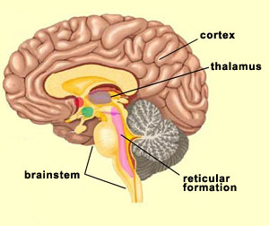
Three large areas of the brain seem
to be especially involved in the phenomenon of consciousness.
The first is the reticular formation,
whose activity level influences the states of alertness, wakefulness,
and sleep. Second is the thalamus, which
sorts the information from the rest of the body and routes
it to other parts of the brain. And finally there is the cortex,
which is of crucial importance for all forms of perception
and all control of voluntary
movements.
Thanks to modern brain-imaging
technologies (follow the Tool Module link to the left), we can
also see the steps that lead to the emergence of a conscious mental
image. For example, which parts of the brain must become active
first, and which ones subsequently, in order for you to have a
conscious visual perception?
To answer this question, neuroscientists
Claire Sergent, Sylvain Baillet, and Stanislas
Dehaene successfully monitored the sequences of neural activity
that occur in a subject’s brain a) when a word briefly projected
on a screen is perceived consciously, and b) when it is not. Whether
the subject perceives the word consciously depends on how long
it is projected. If it is projected for only about a quarter of
a second, it will not be perceived consciously, but if it is projected
for longer—say about three-quarters of a second— it
will.
What do scientists see happening
in the brain when the word is projected for the shorter interval
and for the longer one? Whether or not the word is ultimately
perceived consciously, what happens during the first 275
milliseconds (ms) is exactly the same: only the visual cortex is
activated. (This fits quite nicely with the well
known modular processing in the visual cortex.) But after
that, the brain activity differs according to whether or
not the subject reports having consciously seen the word.
As the animation to the left shows, when the subject does
see the word consciously, the activation is broadly amplified
and reverberates, first through the frontal cortex (starting
after the first 275 ms), then through the prefrontal, anterior
cingulate, and parietal cortexes
(starting after 300, 430, and 575 ms, respectively). But
when the subject does not see the word consciously, the activity
remains localized in the visual cortex and
gradually subsides until it ceases completely after 300 ms.
It thus seems that for consciousness to exist,
there must be some communication or resonance among various parts
of the brain. As we have seen, conscious phenomena do not emerge
from a single location in the brain; instead, they are the product
of a system
involving multiple areas of the brain. That is why, for instance,
when someone’s brain suffers localized damage, their consciousness
may be modified, but rarely eliminated completely.
Another condition for consciousness seems
to be that it can arise only when the “higher” areas
of the brain, such as the frontal cortex, which is connected to
the circuits for emotions and decision-making, are activated.
Forward
of the frontal lobes in the human brain
lie the prefrontal
lobes, which receive countless connections
from other parts of the brain. To cite just two examples,
the ventral
and dorsal visual pathways, which arise
from the temporal and parietal lobes, send projections to
the prefrontal lobes.
The role of the prefrontal cortex is
hard to define clearly, but it seems to be involved in determining
the time sequence
required for a given action. For example,
when people who have damage to the prefrontal cortex are
asked to reproduce a series of movements, they tend to produce
the right movements, but in the wrong order. Also, in tests
where such people are asked to demonstrate various uses for
a given object, they display a great deal of rigidity in
their behaviour and tend to show only the object’s
most common use repeatedly. It is as if they were having
trouble in inhibiting their knowledge of this most common
use so that their knowledge of other uses could emerge.
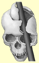
Such
people with damaged prefrontal lobes, as in the famous
case of Phineas Gage (see figure opposite
and links below), may also respond in a stereotyped
way to the sight of an object, even if the social context
makes that inappropriate. For instance, at the sight
of a toothbrush, they might pick it up and starting
brushing their teeth, even if they were in someone
else’s home and the toothbrush weren’t
theirs. When it is pointed out to them that their behaviour
is out of place, they become confused or simply
invent a story that justifies their behaviour.
Because people who have a prefrontal-lobe
deficit are thus at the mercy of the slightest environmental
triggers, they have problems with making plans and
carrying them out. They may thus have some trouble
in retrieving memories if they would need to plan and
apply a search strategy to do so. Two other traits
that such people very often display are a lack of spontaneity
and a fair amount of indifference toward themselves
and others. But despite all this, their general intelligence
remains intact, so they can answer theoretical and
factual questions correctly, but will rarely initiate
a conversation or ask for information.
On September 13, 1848,
Phineas Gage, an American railroad worker, was injured
in an explosion in which an iron rod passed through
his brain. Against all expectations, he recovered
from this accident, but his behaviour was radically
altered. By studying his injuries, scientists gained
a better understanding of the functions of the frontal
lobe. Source: Joan M.K. Tycko