If someone's motor cortex is destroyed
(by a stroke, for example), he or she loses the ability to make precise movements,
especially of the hands and fingers. Learning of new movements is not strongly
affected by damage to the cerebral cortex. The memory of motor sequences learned
previously is also largely spared, though these movements will be executed more
clumsily. These observations show that it is the cerebellum
rather than the cortex that plays an important role in learning and remembering
of movements, also known as procedural
memory.
Area 4 of the precentral gyrus is
not the only area in the cortex that contributes to the pyramidal system. But
is the one where movements can be successfully triggered by lower-intensity electrical
stimuli. In other words, electrical stimuli that are insufficient to produce movements
when applied to other areas of the cortex are sufficient to do so when applied
to Area 4.
THE MOTOR CORTEX
So many different structures
in the brain are involved in motor functions that some people even say that practically
the entire brain contributes to body movements. Though the motor cortex is usually
associated with Areas
4 and 6, the control of voluntary movements actually involves almost all areas
of the neocortex.
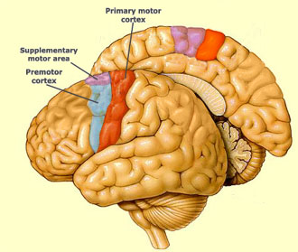
The
primary motor cortex is the anatomical region composed of Area 4 of the precentral
gyrus. Its location was confirmed in the mid-20th century in brain operations
performed by neurosurgeons such as Dr. Wilder Penfield, in Montreal. While performing
operations to alleviate patients' epileptic symptoms, Penfield stimulated various
areas of the cortex to identify vital ones that should not be removed. In this
process, he discovered that stimulations applied to the precentral gyrus triggered
highly localized muscle contractions on the contralateral
side of the body and that there was a somatotopic representation of the corresponding
parts of the body in Area 4 in the primary motor cortex (see box below) .
Penfield also showed that cortical Area 6, just
rostral to Area 4, has two other somatotopic representations that induce complex
movements when stimulated. The first is in the lateral portion of Area 6 and is
called the premotor area (PMA). It helps to guide body movements
by integrating sensory information, and it controls the muscles that are closest
to the body's main axis.
The second somatotopic representation is in the supplementary
motor area (SMA), in the medial part of Area
6. The SMA is involved in planning complex movements and in co-ordinating
movements involving both hands.
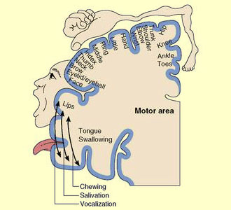
Dr. Penfield's experiments in stimulating
the cortex enabled him to develop a complete map of the motor cortex, known as
the motor homunculus (there are also other kinds, such as the
sensory homunculus). The most striking aspect of this map is that the areas assigned
to various body parts on the cortex are proportional not to their size, but rather
to the complexity of the movements that they can perform. Hence, the areas for
the hand and face are especially large compared with those for the rest of the
body. This is no surprise, because the speed and dexterity of human hand and mouth
movements are precisely what give us two of our most distinctly human faculties:
the ability to use tools and the ability to speak.
The functions of the basal ganglia
are complex and still largely unknown. People who have Parkinson's disease, characterized
by trembling and by difficulty in initiating movements, show a deficiency of dopamine
in their basal ganglia. Because these structures play an important role in determining
various aspects of movement, their malfunctioning results in the motor problems
associated with Parkinson's disease.
Some abnormalities also are found
in the basal ganglia of people who have Huntington's Disease or Tourette Syndrome.
These patients experience involuntary movements that cause all sorts of grimaces,
tics, and spasms.
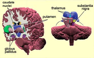
THE BASAL GANGLIA
The term "basal ganglia"
refers to a group of several structures in the brain: the caudate nucleus, the
putamen, the globus pallidus, and the subthalamic nucleus. The substantia nigra,
a midbrain structure that has many interconnections with the basal ganglia, is
not actually part of this grouping but is often associated with it.
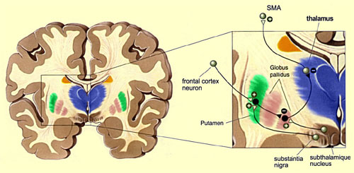
The basal ganglia are involved in a complex
loop that connects them to various areas of the cortex. The information from
the frontal, prefrontal, and parietal areas of the cortex passes through the basal
ganglia, then returns to the supplementary motor area via the thalamus. The basal
ganglia are thus thought to facilitate movement by channelling information from
various regions of the cortex to the SMA. The basal ganglia may also act as a
filter, blocking the execution of movements that are unsuited to the situation.
Not all of the circuits involving the basal ganglia are motor circuits, however.
Many are instead involved in memorizing and in cognitive and emotional
processing. A great deal about the basal ganglia remains unknown. They seem to
play a far larger role than just their contribution to motor control.
The cerebellum also acts as a learning
and memorizing machine, thanks to its modifiable
neural connections that continuously compare everything they are programmed to
do with the results that they are actually achieving. When this comparison does
not allow the expected result to be achieved satisfactorily, the cerebellum's
activity modifies the sequence of movements in a compensatory manner to make them
more effective. This procedural
memory thus develops automatically with practice, without the help
of any conscious control.
The cerebellum also appears to play
a major role in learning how to co-ordinate the various segments of the body.
The movement of each segment of your body affects the next, because of its mass.
The cerebellum therefore apparently learns how to calibrate its commands to the
muscles in terms of strength and duration in order to correct in advance for the
effects of these interactions along the path of motion.
THE CEREBELLUM
The
cerebellum appears to play several roles. It stores learned sequences of movements,
it participates in fine tuning and co-ordination of movements produced elsewhere
in the brain, and it integrates all of these things to produce movements so fluid
and harmonious that we are not even aware of them.
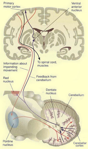
To
do all this, the cerebellum maintains close communications with the cortex. The
motor, somatosensory, and posterior parietal areas of the cortex project massive
numbers of axons to the nuclei of the pons, located in the brainstem. The neurons
of the pons then project their axons into the cerebellum. This corticopontocerebellar
tract forms an extremely dense nerve bundle containing about 20 million axons,
just about 20 times more than the pyramidal bundle!
The two hemispheres
of the cerebellum then sends signals back to the motor cortex via interconnections
in the ventrolateral nucleus (VLc) of the thalamus. The cerebellar hemispheres
thus influence the muscles of the arms and legs via the cortex and the lateral
motor system.
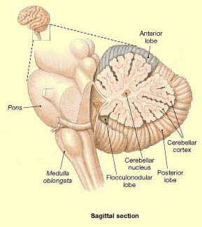
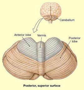
The two hemispheres of the cerebellum are not
divided neatly in two like the two hemispheres of the cerebrum. The medial portion
constitutes what is known as the cerebellar vermis. This vermis does not display
any lateralization. It projects axons to the brainstem which, via the ventromedial
system, help to maintain posture.
The brain mechanisms that go into
planning and executing a movement are far more complex than the motor cortex's
simply issuing a command and the motor neurons' executing it.
For example,
suppose that you go to pick up a glass of water that you think is cool and refreshing,
but is actually boiling hot. As soon as you touch the glass, you pull your hand
back immediately, by reflex,
without thinking about it.
But suppose that next, your child tries to
grab this glass, which you already know is hot. In this case, because your child's
safety is so important to you, you can consciously overcome the reflex to pull
your hand away. Instead, using your voluntary motor control, you grab the glass
yourself and put it where your child can't reach it.
Lastly, if someone
tells you that the glass is made of fine crystal and not ordinary glass, you will
probably handle it more carefully. In other words, your brain will take this information
into account and adapt your method of grasping the glass accordingly.
All of these facts demonstrate that the execution of a movement is not simply
a matter of the brain's sending a "Go!" command to some motor neurons
in the spinal cord, but rather the result of a highly elaborate construct. Moreover.
the remarkable adaptability of motor activity demonstrates the involvement of
powerful regulatory
and feedback mechanisms.
THE ACTIVATION SEQUENCE FOR THE MOTOR AREAS
The information processing
that the brain must perform to initiate a voluntary movement can be divided into
three steps. The first step is to select an appropriate response
to the current situation, out of a repertoire of possible responses. This response,
which corresponds to a particular behavioural objective, is determined in a global,
symbolic fashion.
The second step is to plan the movement in
physical terms. This step consists in defining the characteristics of
the selected response as the sequence of muscle contractions required to carry
it out.
The third step is to actually execute the movement.
It is in this step that the motor neurons are activated that trigger the observable
mechanics of the movement.
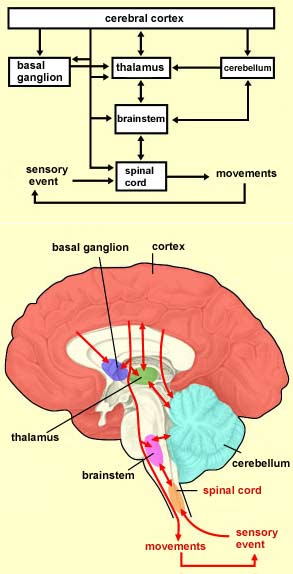
Consequently, the control
messages issued by the motor cortex are themselves triggered by messages from
other
cortical areas. The motor cortex also communicates closely with subcortical
structures such as the basal ganglia
and the cerebellum, through the thalamus,
which acts as a relay.
In light of what we now know about the sequence in
which the motor areas of the cortex are activated, we can deconstruct the classic
sequence "Ready? Set. Go!" in terms of localized activity in the brain.
In the "Ready?" phase, the parietal and frontal lobes become active
first, with a contribution from the subcortical structures involved in vigilance
and attentiveness. The "Set" command then activates the supplementary
and premotor cortical areas, where the strategies for movement are developed and
maintained until the "Go!" signal is given. The "Go!" signal
may come from an outside source, as it does in an actual race, or it may come
from inside the person doing the running, who decides for himself or herself that
all the conditions are present to start running. The "Go!" command then
applies information from subcortical structures such as the basal ganglia that
will influence Area 6, and then eventually the primary cortex, which will
cause the action to be carried out.