Funding for this site is provided by readers like you.
Pleasure and pain
Sub-Topics
Pleasure-Seeking Behaviour
Pleasure and Drugs
Avoiding Pain

Linked
HelpLink : LE CONTROLE DE LA DOULEURLink : http://neurobranches.chez.tiscali.fr/systnerv/systsens/douleur1.htmlLink : http://www.lib.mcg.edu/edu/eshuphysio/program/section8/8ch5/s8ch5_45.htm
Link : Pain PhysiologyLink : Mechanisms of Disease: neuropathic pain-a clinical perspectiveLink : SOMATOSENSORY PATHWAYS FROM THE BODY


The phylogenetic classification of the pain pathways is not the only one. The nociceptive pathways can also be identified simply according to the routes that they follow anatomically. Thus the archispinothalamic and paleospinothalamic pathways, both of which leave many collaterals in the reticular formation of the brainstem, are also referred to as spinal-reticular-thalamic pathways.


When ascending pain signals cross the medulla oblongata, they can activate certain neural pathways of the autonomic nervous system that then cause increases in heart rate, respiratory rate, blood pressure, perspiration, and so on. The size of these increases depends on the intensity of the pain and can be reduced by the descending control pathways originating in the higher centres of the brain.


Pain and temperature signals from the face and the rest of the head are transmitted to the brain by a system that is similar to but distinct from the one in the spinal cord. This system is composed of the trigeminal pathways.

In these pathways, the cell bodies of the first sensory neurons are located in the trigeminal ganglion and in other ganglia associated with cranial nerves VII, IX, and X. The axons of the trigeminal nerve (cranial nerve V) then enter the pons and descend into the medulla oblongata, where they synapse with the second neurons in the circuit. As in the spinal cord, the axons of the second neuron cross the midline and go up the contralateral side of the body to the thalamus, where they connect into the ventral posterior medial nucleus of the thalamus, a more medial part of the thalamus than in the spinothalamic pathway.

Lien : Noyaux des nerfs crâniens dans le tronc cérébralLien : Nerf trijumeauLien : Trigeminal nerve nuclei
ASCENDING PAIN PATHWAYS
DESCENDING PAIN-CONTROL PATHWAYS

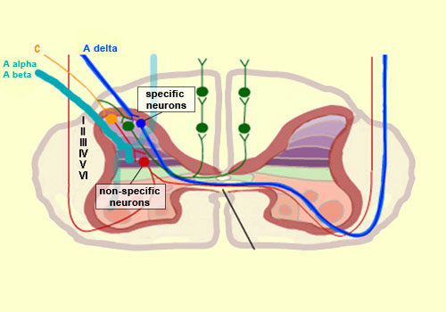
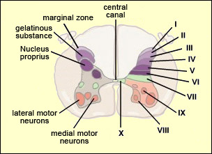
From the reception of a pain stimulus in the peripheral nervous system to the perception of pain in the brain, which generates various behaviours in response, the pain circuit follows several different, often redundant pathways. This is not surprising, considering how important pain is for the body’s integrity.

These multiple nociceptive pathways all start in the same way. A pain signal coming from the skin, for example, first travels up a sensory nerve fibre composed of the axons of the T-shaped sensory neurons located in a spinal ganglion. These axons then enter the spinal cord, where they immediately divide and travel a short distance of one or two segments upward and downward in the spinal cord. They thus form what is called Lissauer’s tract before terminating in the external part of the dorsal horn.

The particular areas of the dorsal horn where the various fibres (A alpha, A beta, A delta and C) synapse are not random: they map to a very specific spatial organization.

Thus the large-diameter, myelinated fibres (A alpha for touch and A beta for proprioception), which ascend directly into the ipsilateral dorsal column of the spinal cord, nevertheless also send out collateral axons that penetrate into the deepest layers of the dorsal horn (see box below), which make direct contact with the deep layers of the ventral horn. There the connections are made with the motor neurons that make the withdrawal reflex possible.

Meanwhile, the smaller-diameter A delta and C fibres, which transmit pain, synapse on two main types of neurons that are also located in specific layers of the dorsal root of the spinal cord.

The first type are known as specific nociceptive neurons, and their cell bodies are located in layers I and II. These neurons receive A delta and C fibres and hence are activated exclusively by mechanical and/or thermal nociceptive stimuli applied to the skin. The axons of these specific nociceptive neurons combine to form the neospinothalamic tract.

The second type are known as non-specific neurons, and their cell bodies are located in layer V. These neurons respond preferentially but not exclusively to nociceptive stimuli. They are also known as convergent, polymodal, or wide-dynamic-range neurons, because they can be activated by fibres carrying non-nociceptive mechanical stimuli as well as by pain stimuli of tactile, muscular, or visceral origin. These neurons can also encode the intensity of the peripheral stimulus, by increasing their firing frequency as this intensity increases. Once this frequency exceeds a certain threshold, the message becomes nociceptive.

The receiving field on the skin from which these non-specific neurons can be activated is larger than the field for specific neurons and also displays a sensitivity gradient: at the centre of the receiving field, all mechanical stimuli produce a nerve impulse, while at the edge, only nociceptive stimuli do so.

The nerve impulses observed in non-specific neurons can therefore have an early component, due to the activation of the collaterals of the A alpha and A beta fibres, and then later components due to the activation of the A delta and C fibres in response to more intense stimuli.

The fact that fibres of various diameters and of both visceral and tactile origin converge on these neurons also explains two distinct phenomena: projected pain, in which a pain in the internal organs is felt in an area of the skin, and the segmental control of pain by the activation of non-nociceptive afferent pathways.

The axons of the non-specific nociceptive neurons form the paleospinothalamic tract, which is of earlier evolutionary origin than the neospinothalamic tract formed by the specific neurons described earlier. And both of these tracts are newer than the archispinothalamic tract, which is of very early origin.

But all of these tracts are formed by the axons of the second principal neuron in the nociceptive circuit leading to the brain (the blue neuron with the blue number 2 in the diagram to the right). This neuron is located in the dorsal horn of the spinal cord. The expression “principal neuron” is used here, because the dorsal horn also contains many interneurons that modulate the transmission of pain impulses over this circuit.

Adapted from: Neuroscience, 3rd Edition, Purves, Augustine, Fitzpatrick, Katz, LaMantia, McNamara, Williams, De Boeck, Eds., 2003.

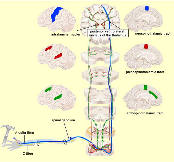
Thus pain signals are carried over three main extralemniscal pathways (as opposed to the lemniscal pathway, discussed below) that appeared successively in the course of evolution: the archispinothalamic tract, the paleospinothalamic tract, and the neospinothalamic tract.

For an animated version of this figure, see http://neuroscience.uth.tmc.edu/s2/ii7-2.html.


The archispinothalamic tract or archispinothalamic pathway is the oldest pathway that pain stimuli can take to reach the brain. It is a multisynaptic, diffuse pathway. The first places that it reaches in the brain are the reticular formation and the periaqueductal grey matter, followed by the intralaminar nuclei of the thalamus. This tract also sends collaterals to the hypothalamus and to various nuclei in the limbic system, thereby contributing to visceral, autonomic, and emotional reactions to pain.


The pathway that appeared next in the course of evolution is the paleospinothalamic tract or paleospinothalamic pathway. (Note that this phylogenetic or evolution-based classification is not the only possible one—see first sidebar.) The paleospinothalamic tract is composed of small-diameter fibres that conduct nerve impulses slowly. Like the archispinothalamic tract, it has no somatotopic organization. It projects widely into the reticular formation at all levels of the brainstem, thus contributing to two important phenomena. The first is the maintaining of wakefulness in the central nervous system by the ascending reticular system. The second is the activation of certain nuclei in the brainstem that constitute the start of the descending pain-control pathways.

The terminations of the paleospinothalamic pathway also continue into the intralaminar nuclei of the thalamus. The neurons of these nuclei (which are the third neurons in the afferent nociceptive pathways, after the neurons in the spinal ganglia and the neurons in the dorsal horn of the spinal cord) send projections into various cortical regions, including the frontal cortex, cingulate cortex, and insular cortex.


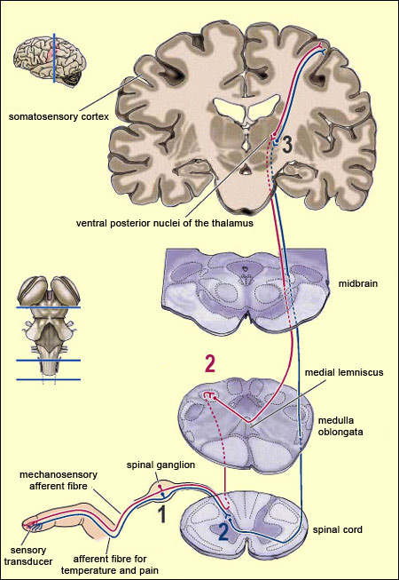
The neospinothalamic pathway or neospinothalamic tract, usually referred to simply as the spinothalamic pathway, is the most recent nociceptive pathway from an evolutionary standpoint and is found in higher mammals only. This is the pathway travelled by the rapid component of pain, which tells the brain about the nature of the painful stimulus (sting, burn, etc.) and its precise bodily location . This is also the pathway that transmits the sensation of temperature.

The spinothalamic pathway consists of the axons of the specific neurons of the ventral horn of the spinal cord. All of these axons decussate, that is, cross over to the contralateral side of the spinal cord. (They thus differ from the axons in the archispinothalamic and paleospinothalamic pathways, which make bilateral connections to the brain structures that they innervate, because some of their collaterals do not decussate and instead ascend directly on the same side.)

Once they decussate, the axons of the spinothalamic pathway continue their ascent on the contralateral side, through the anterior lateral part of the spinal cord (which is why this pathway is also sometimes called the anterolateral or ventrolateral tract). The axons then enter the medulla oblongata, where they are joined by the axons of the trigeminal spinal nucleus, which convey pain sensations from the face (see sidebar), as well as by the medial lemniscal pathway, which is responsible for the sense of touch.

Most of the fibres in the neospinothalamic tract that come from parts of the body below the neck terminate in the posterior ventrolateral nucleus of the thalamus. The neurons in this nucleus are the third in the neospinothalamic pathway and send their axons to the primary somatosensory cortex.

The dorsal and ventral horns of the spinal cord contain 10 different layers of grey matter, known as the Rexed laminae. Just as the Brodmann areas map the cortex according to the cytoarchitecture (cellular structure) of each of its parts, the Rexed laminae distinguish 10 different layers in the spinal cord on the basis of the characteristics of their neurons.

The dorsal horn, where the first connections in the pain pathways are made, contains laminae I to VI, while the ventral horn, comprising the motor neurons, contains laminae VII to IX. Lamina X surrounds the central canal.

Numerous studies of the physiology of the spinal cord have confirmed that this division based on cell types has functional bases as well.

Lien : Internal Structure of the Spinal CordLien : Rexed laminaeOutil : Les aires corticales de Brodmann


Linked
Link : Animation: A novel mechanism of allopathic pain, by Michael S.C. CorrinLink : How Pain WorksLink : Neuroanatomy of PainLink : L'effet placebo livre ses secrets
Link : La douleurLink : Bases physiopathologiques et évaluation de la douleurLink : NociceptionLink : Informations sur la fibromyalgie
Link : Synthèse des mécanismes impliqués dans un syndrome douloureux complexe : la fibromyalgieLink : Connectivity of the human periventricular-periaqueductal gray regionLink : Bases neurophysiologiques du phénomène de contre-irritation : les contrôles inhibiteurs diffus induits par stimulations nociceptives
Experiment
Experiment : Activation of the Opioidergic Descending Pain Control System Underlies Placebo AnalgesiaExperiment : Lesion and electrical stimulation of the ventral tegmental area modify persistent nociceptive behavior in the ratExperiment : A role for peripheral somatostatin receptors in counter-irritation-induced analgesia


The reduced levels of norepinephrine and serotonin observed in people who have fibromyalgia suggest that a deficit in the diffuse noxious inhibitory controls (DNICs) may be the cause of this painful syndrome. For example, the raphe nuclei, which are involved in DNICs, use serotonin as a neurotransmitter. This would explain why people with fibromyalgia experience relief from treatments such as tricyclic antidepressants and aerobic exercise.

It is also believed that acupuncture may achieve some of its analgesic effects by bringing DNICs into play.


Much of what we know about the central mechanisms of pain control comes from experiments in which electrical and pharmacological stimuli were applied to certain areas of rat and human brains.

When an electrode is implanted in and used to apply an electrical stimulus to the periaqueductal grey matter of a rat’s brain, the initial effect, as determined from the rat’s behaviour, is analgesic. This electrical stimulus may also reduce the activity that a nociceptive stimulus normally generates in the neurons of the dorsal horn of the spinal cord. But this same stimulus has no effect on the rat’s sense of touch or sensitivity to temperature.

Neurosurgeons have successfully relieved severe pain in human patients by means of electrodes implanted close to the periaqueductal grey matter in their brains. This technique, known as deep brain stimulation (DBS), has been used since the 1980s to treat recalcitrant pain.

Researchers suspected very early on that this electrical stimulation might produce this analgesia by causing the release of endorphins, in part because administering naloxone, an opiate antagonist, blocked this analgesic effect.

More direct evidence was obtained when local microinjections of morphine into the periaqueductal grey matter were found to produce a strong analgesic effect. This microinjection method enabled researchers to identify several areas of the medulla oblongata that are involved in suppressing pain, such as the nucleus raphe magnus, the giganto-cellular nucleus, and the lateral reticular nucleus of the solitary tract. But depending on which part of the periaqueductal grey matter received the electrical stimulus, naloxone did not always block the analgesia, which indicated that the descending pain-control pathways utilize several other neurotransmitters besides opioids. For example, neuroscientists now have extensive knowledge of the serotonergic pathways descending from the raphe nuclei and have learned that serotonin antagonists can also cancel out the analgesic effect produced by electrical stimulation of the brain.

Lien : Transcutaneous electrical nerve stimulationLien : Deep brain stimulationLien : Stimulation cérébrale profondeExpérience : Analgesia from Electrical Stimulation in the Brainstem of the Rat
DESCENDING PAIN-CONTROL PATHWAYS
ASCENDING PAIN PATHWAYS

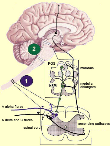
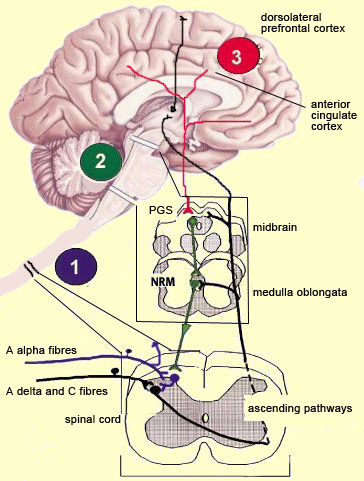
The brain is not a passive receiver of sensory messages, but rather a centre that interprets them and makes constant adjustments accordingly. For example, everyone knows that the way you perceive pain will be influenced by whether you focus on it or think of something else instead. And it seems reasonable to suppose that evolutionary selection may have favoured those individuals who could ignore pain signals for long enough to take actions that let them escape and survive danger.

What interests neuroscientists is just how people manage to control and attenuate their perception of pain. The mechanisms for the descending control of pain were first described in detail in relation to the spinal cord, by Melzack and Wall’s gate theory. This theory marked the end of the conception of pain as a simple, primitive alarm system, by showing that the transmission of pain signals was controlled in some ways in the spinal cord and hence possibly at various locations in the brain as well. Thus the eminently subject nature of pain, and the many psychological factors that can affect it, were found to have a neural substrate.

The neural pathways originating in the higher brain centres that generate these psychological states exert their influence on subcortical areas and are hence by definition descending pathways. These pathways have an inhibitory effect that, depending on how much it is activated, can “close the gates” to various degrees in the centres that relay pain signals up the ascending nociceptive pathways.


For example, the diffuse noxious inhibitory controls induced by pain stimuli originate in the brainstem. These control mechanisms are often referred to by their acronym, DNICs, and scientists have known about them for a long time, because they are the means by which one pain masks another. In ancient times, burning-hot metal tips were applied to various points on patients’ bodies to relieve certain kinds of pain elsewhere. In modern times, when cattle ranchers need to carry out painful procedures such as castration, they may apply a clamp to the animals’s nose to mask the pain. In other words, when two pain stimuli are applied to two different areas of the body that are distant from each other, perception of the weaker of the two stimuli is inhibited.

In the late 1970s, a formal hypothesis was developed to explain this phenomenon: specifically, that a localized nociceptive stimulus can result in a generalized inhibition of afferent nociceptive nerves elsewhere in the body. For example, researchers have conducted experiments in which they applied a pain stimulus to the tip of a rat’s paw and then monitored the electrical activity in a nociceptive neuron in the spinal cord that was responding to this stimulus. The researchers then found that they could inhibit this nociceptive response by applying pain stimuli to numerous points elsewhere on the rat’s body. The researchers also found that non-nociceptive stimuli were totally ineffective in this regard, and that the degree of inhibition was proportional to the intensity and duration of the pain stimulus applied to inhibit the nociceptive response.

In the model proposed by Le Bars et al., DNICs facilitate the detection of nociceptive messages by reducing the activity of converging neurons that are not concerned with this pain. The greater contrast thus created between the field of the activated neuron and the silencing of the unconcerned neurons would thus enable the brain to better identify the precise location of this pain.

This general silencing is based on the fact that ascending nociceptive nerves not only transmit pain signals to the higher centres in the brain, but also make connections to the midbrain and the brainstem, more specifically in the periaqueductal grey matter (PGM) and in the nucleus raphe magnus (NRM). These structures then return efferent signals downward to various levels of the spinal cord, where, with the help of inhibitory interneurons, they produce a diffuse inhibition.

Green: diffuse inhibitory controls induced by nociceptive stimuli
Violet: segmental controls of non-pain peripheral origin


Now we will discuss the descending controls of supraspinal (central) origin that are associated with more complex psychological phenomena, such as hypnosis and the placebo response, that can reduce our perception of pain to varying degrees.

In this case, the activation sequence for the descending pathways appears to involve brain structures such as the dorsolateral prefrontal cortex (an area involved in predictions based on beliefs) which, through synaptic connections using endorphins, communicates with the anterior cingulate cortex. This structure would then in turn activate the periaqueductal grey matter, and then the raphe nuclei and other nuclei in the brainstem. Complex modulations occur at each of these sites, in addition to those that will occur in the dorsal horn of the spinal cord much farther down.

Red: descending controls of supraspinal (central) origin, associated with psychological factors
Green: diffuse inhibitory controls induced by nociceptive stimuli
Violet: segmental controls of non-pain peripheral origin

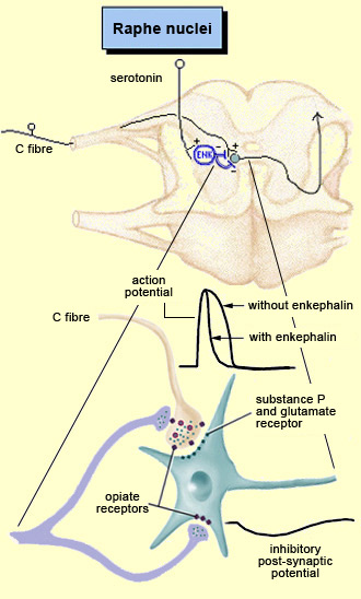
The periventricular grey matter and its continuation, the periaqueductal grey matter, which is located around the cerebral aqueduct in the midbrain, have been studied extensively (see sidebar). It is now known that both structures not only send descending projections to the spinal cord and the cerebellum, but also send ascending projections to the thalamus and the frontal lobes. Neuroscientists therefore believe that this periventricular and periaqueductal area may modulate pain both centrally and in the spinal cord.

There are, however, very few projections from the periaqueductal grey matter directly to the spine, and this grey matter is believed to exert its effect primarily through the serotonergic raphe neurons. Noradrenergic neurons in the locus coeruleus and dopaminergic neurons in the ventral tegmental area also appear to be involved.

The axons of all these neurons join to form a bundle of descending fibres, the dorsolateral funiculus, that terminate on the inhibitory interneurons in the dorsal horn of the spinal cord. Thus these interneurons, in addition to being subject to the segmental influence of peripheral origin, may also be subject to an activation of central origin.

The interneurons in the dorsal horn exert their inhibitory effect by releasing endogenous opioids that act on specific receptors located both in the terminal buttons of the primary nociceptive afferents and directly on the converging neurons. Because the inhibition is thus exerted both pre- and post-synaptically, it is doubly effective: it reduces the strength of the nociceptive message transmitted to the next neuron, while also hyperpolarizing that neuron and thus reducing the probability that it will transmit action potentials in turn.

The interneurons (violet) use the neurotransmitter enkephalin to inhibit the projection neuron (green) in two ways.


  Presentations | Credits | Contact | Copyleft