Funding for this site is provided by readers like you.
Body movement and the brain
Sub-Topics

Making a Voluntary Movement


Linked
Help Link : Pyramidal Motor System Corticospinal tract Link : Le mouvement
Experiment
Experience : Knee Jerk Reflex (Patellar Reflex)

Stephen Hawking, the famous astrophysicist, is confined to a wheelchair because of amyotrophic lateral sclerosis, a degenerative disease of the motor neurons.

Link : Stephen Hawkins: my experience with ALS Link : The Motor Neurone Disease (MND) Association’s Link : International Alliance of ALS/MND Associationson the internet Link : The ALS Society of Canada

Because nerve impulses must travel relatively large distances down the axons of the pyramidal neurons and motor neurons (the longest of which are over a metre long!), nature has devised two ways of making these impulses travel faster. One way is by making some axons slightly larger in diameter, which reduces resistance to these electrical currents. The other way is by dividing most axons into a series of small segments each of which is wrapped in an insulating sheath known as myelin. The nerve impulses are thus forced to "jump" each gap between myelin segments, and this arrangement too increases the speed at which the impulses are conducted.

THE AXONS ENTERING AND LEAVING THE MOTOR CORTEX
THE DISTINCTIVE CELL ARCHITECTURE OF THE CEREBELLUM

In the brain, there are never any dead ends. Every nucleus or group of neurons receives connections from a multitude of neurons and sends axons to other parts of the central nervous system. The motor cortex is no exception.

The axons entering the motor cortex come mainly from neighbouring regions of the cortex as well as from the thalamus. The axons leaving the motor cortex come from the largest cells in the cortex, known as the pyramidal neurons, some of which are almost 0.1 mm in diameter!. These axons are extremely long, because they descend all the way down into the spinal cord, where they make connections with the motor neurons located there.

Thus, from your brain to your muscles, it takes just two neurons to relay the command for a voluntary movement: one of the pyramidal neurons, whose axons are bundled into various tracts that descend to the motor neurons in the spinal cord, and one of these motor neurons, whose axons emerge from the spinal cord to form the motor nerves that excite your muscles and produce their movements.


The voluntary movements thus produced as the result of commands originating in various parts of your brain enable you to adapt to various situations. In contrast, reflex movements are involuntary, simple, rapid, and stereotyped. The circuits that enable reflex movements are often located in the spinal cord and, unlike voluntary movements, do not require any commands from the brain.


Like most brain functions, motor controls are crossed: the right motor cortex controls the left side of the body, and the left motor cortex controls the right side. The axons of the neurons in each of these cortexes must therefore bifurcate (split in two) somewhere during their descent to the spinal cord so that they can change sides. This crossover, or decussation, occurs just before the junction between the medulla oblongata and the spinal cord.

This decussation of the pyramidal tract is the reason that brain injuries and strokes on one side of the head typically cause paralysis on the other side of the body.


Sometimes, if you let the weight of your body press on one of your arms or legs for too long, it will become numb. This disagreeable feeling of not being able to feel or move your arm or leg occurs when certain motor nerves become pinched. The nerve impulses are then blocked in both directions. The sensory impulses from your arm or leg cannot reach your spinal cord, and the motor impulses from your brain cannot reach your muscles. When the pressure on the nerve is released, all the nerve impulses start moving again once. This sudden recovery confuses your brain slightly, causing it to interpret the impulses as pain. That is the reason for the intense feeling of "pins and needles" that follows.


THE DISTINCTIVE CELL ARCHITECTURE OF THE CEREBELLUM
THE AXONS ENTERING AND LEAVING THE MOTOR CORTEX

The cellular structure of the cerebellum (meaning "little brain") is similar to that of the telencephalon in the cerebrum. The grey matter (containing the cell bodies of the neurons) is found at two locations. One is at the surface of the cerebellum, in a thin layer called the cerebellar cortex (the cerebellum's "bark", so to speak). The other is deep inside the cerebellum, where neurons are grouped in clusters called cerebellar nuclei.

The cerebellar cortex contains many furrows, running mainly in a transverse direction. The deepest furrows divide the cerebellum into lobules. The shallower furrows within each lobule divide it into lamellae.

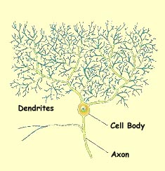
The main cells of the cerebellar cortex are large, pear-shaped cells called Purkinje cells, after the Czech anatomist who first described them, in 1837. These cells receive impulses from their synapses with the afferent nerve fibres entering the cerebellum, then send these impulses out along their axons to the cerebellar nuclei. The Purkinje cells form a layer parallel to the surface of the cerebellar cortex. There are also two other layers of neurons on either side of the Purkinje cells.

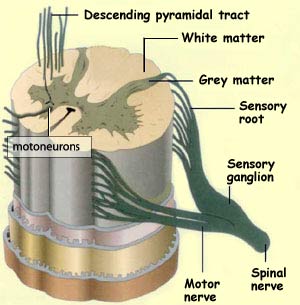
As in the cerebral cortex, the white matter that forms the interior of the cerebellum consists of the myelinated nerve fibres that come and go between the cerebellar cortex , the nuclei deep inside the cerebellum, and the other brain structures outside it.


  Presentations | Credits | Contact | Copyleft