| |


The mirror neurons in the premotor area
of our brains do not react to just any movements of someone else’s mouth
or hands, but only
to those movements that are goal-oriented. The response of these neurons
thus depends on the goal that we discern in the observed movement—the intention
that we attribute to it. The mirror neurons in our brain’s motor
system can thus help us to decode the meanings of actions performed by other people,
and hence their
mental states. Some authors think that this mechanism for interpreting
gestures is
also applied to verbal communication, in particular in the perception of speech.
|
People who are mourning a death or a lost love
or reacting to some form of social rejection often describe their emotional
pain in terms similar to those used for physical pain. According to
many pain specialists, the connection between the two is more than metaphorical.
For example, in a study where the subjects played a video game that made
them feel rejected, brain images revealed activity in the anterior
cingulate cortex, a part of the brain that is important in the physical-pain
matrix—the brain circuits that let us know the risks of being injured
or that lead us to take special care of parts of our body that have in fact been
injured. Because we are a highly social species in which mutual
assistance is important, becoming isolated or losing our most significant
personal relationships has always constituted a threat to our survival, whether
by making us more vulnerable to predators in prehistoric times or by causing us
to join the ranks of the socially outcast and homeless in today’s big cities.
Thus our social-attachment system seems to use our physical-pain
system to ensure that we maintain relations with other people. Accordingly, being
separated from a loved one or rejected by a group is painful to us, and we tend
to avoid such situations when we can. | | |
| SHARING OTHER PEOPLE’S PAIN | |
Our
ability to feel what other people are feeling seems to be manifested through
mechanisms that are largely involuntary and non-intentional. Thus, human empathic
responses appear very early in
the course of children’s development; children seem to be literally
“pre-wired” to form
relationships with their mothers. This
affective resonance is important not only for creating the critical bond between
children and their mothers, but also for creating the emotional ties with other
individuals that will foster mutual
assistance and reproduction
later in life, and hence the survival of the species in the long run. This automatic
mental resonance with other people also helps us to make quick predictions about
other people’s actions and needs, thereby improving communication, an important
adaptive asset in a social species such as ours. Human
empathy would thus appear to be deeply rooted in the evolutionary
history of our non-human primate ancestors and, many authors believe, would
appear to provide a biological basis for our moral behaviours. Scientists have
proposed many possible mechanisms to explain how human empathy may have developed.
Among the most significant of these mechanisms are those involving mirror neurons. The
first part of the brain in which mirror neurons were identified was the premotor
cortex, in the early 1990s. Some neurons in this area become activated when
we make a particular motion, such as picking up a cup to drink from it. However,
some of these same neurons also become activated when we are completely motionless
ourselves but see someone else making exactly the same motion. The discovery that
a visual stimulus can thus activate neurons in a motor area of the brain in a
very specific way aroused great interest in the scientific community. Subsequent
studies identified mirror neurons in various other parts of the brain as well.
Because mirror neurons let us simulate in our own brains what is going on in someone
else’s, this combination of their neurons and our own has been described
as a “shared neural network”. Studies have found such shared neural
networks not only in the sensorimotor circuits of the brain, but also in the brain
circuits responsible for emotional reactions. This may help to explain why, for
example, we can be moved to tears by an actor in a film or on stage: the actor’s
expressions of an emotion may activate the parts of our own brains that correspond
to this emotion. These findings made
it necessary to define mirror neurons more broadly as a class of neurons that
are activated not only when we experience an endogenous cognitive event ourselves,
but also when we see signs that another individual is experiencing (or is about
to experience) the same kind of cognitive event.

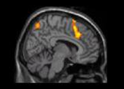
The top picture, of one person’s finger in a painful situation, activates
the anterior cingulate cortex (the larger of the two yellow areas in the bottom
picture) in the brain of a person who is looking at it.
Source: Jean
Decety, University of Washington, Seattle
| | In
the case of pain, for example, one such sign might be that person’s behaving
in some way that expresses suffering, or simply making a facial expression indicating
that he or she is in pain. But the discharge of our mirror neurons associated
with pain could also be triggered simply by the sight of a knife being pressed
against someone else’s skin (not even necessarily someone we know). A
number of brain-imaging studies have located a network
of areas in the brain that are active when we experience physical pain. These
studies have also shown, however, that when we have an empathic response to someone
else’s pain, not all parts of these same circuits become active, but mainly
the parts associated with the unpleasant affective component of pain: the anterior
cingulate cortex and the anterior insula. The primary and secondary somatosensory
areas, as well as areas such as the posterior insula, are highly active when we
are experiencing pain ourselves, but do not react as much when we observe someone
else’s pain. | These
studies thus reveal a partial overlap between someone’s neural activity
when they are actually having a painful experience themselves and their neural
activity when they are observing someone else having such an experience. The partial
nature of this activation has
suggested some possible explanations of how we distinguish these two situations
so as not to confuse someone else’s distress with our own.
Philip L. Jackson and other authors have shown that experiencing empathy for other
people’s pain does not generate the same activation patterns in our brains
as imagining ourselves to be in pain when we are not actually in the midst of
a painful experience. When we imagine ourselves in pain, the pain matrix is activated
in far broader areas of the brain, including in particular the secondary somatosensory
cortex and the dorsal portion of the anterior cingulate cortex. However,
some studies using transcranial magnetic stimulation have shown that we do have
some ability to map the part of another person’s body that is in pain onto
our own somatosensory cortex. Further experiments will be needed to determine
the true extent of this isomorphism in the empathic response. Be
that as it may, the role of brain structures such as the anterior cingulate cortex
in empathic responses to other people’s observed emotions has been confirmed
in situations not involving pain. For example, this structure becomes activated
not only when someone smells an unpleasant odour themselves, but also when they
see an expression of disgust on the face of someone else who is smelling such
an odour.
Some individuals lack empathy and the ability
to perceive other people’s emotions. This is the case for psychopaths,
who often have only very superficial affective states, feeling little or no remorse,
for example, when they commit a crime. This difficulty in experiencing their own
emotions makes it hard for them to recognize other people’s distress, as
has been confirmed by the limited responses measured in the autonomic
nervous systems of psychopaths while they are witnessing other people
experiencing fear, sadness, or disgust. People with autism
are another group who often show a limited ability to feel empathy for others.
In one study, for example, Iacoboni asked autistic children and normal children
to observe and imitate facial expressions for various emotions while he recorded
the children’s brain activity with a functional magnetic resonance imaging
device. Though both groups performed the task successfully, the autistic children
showed less activity in their mirror-neuron circuits, especially in the inferior
frontal area, while doing so. Moreover, the extent to which this activity was
reduced corresponded to the severity of their autistic symptoms. Iacoboni therefore
concluded that the integrity of our mirror-neuron systems seems to be essential
for normal social development. |
|
|