In brain imaging, when subjects
are asked to move their thumbs, activity is observed in the posterior parietal
and somatosensory areas, Area 8 of the prefrontal cortex , and motor areas 4 and
6. Interestingly, if subjects are simply asked to repeat
the movement mentally without actually performing it, Area 6 is still
activated, but not Area 4.
Some kinds of damage to the posterior
parietal cortex can lead to a syndrome called apraxia. In one form of apraxia,
patients can make certain gestures spontaneously but have trouble in making these
same gestures if asked to do so. In another form, patients cannot make the correct
movements to use objects such as a pencil or a pair of scissors, even though they
can describe the functions of these objects perfectly.
In addition, people
with apraxia will have even more difficulty in performing a gesture if they are
asked to do so outside the appropriate social context. What apraxia thus seems
to impair is the ability to make voluntary movements that are not directly elicited
or stimulated by the environment.
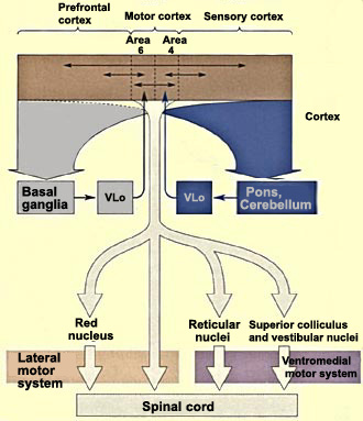
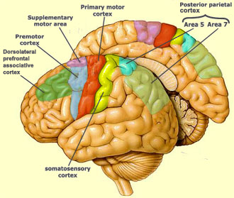
THE MOTOR CORTEX
The anatomical region
of the brain known as Area 4 was given the name primary motor cortex (symbol:
M1) after Penfield
showed that focal stimulations in this region elicited highly localized muscle
contractions at various locations in the body. This mapping is represented somatotopically
on the motor cortex, where the surface area devoted to controlling the movements
of each body part varies in direct proportion to the precision of the movements
that can be made by that part (see boxed text below).
The
motor cortex also includes Area 6, which lies rostrally to Area 4 and is divided
into the premotor area (or premotor cortex) and the supplementary motor area.
The premotor cortex is believed to help regulate posture by dictating an optimal
position to the motor cortex for any given movement. The supplementary motor area,
for its part, seems to influence the planning and initiation of movements on the
basis of past experience. The mere anticipation of a movement triggers neural
transmissions in the supplementary motor area.
Besides
the frontal cortex, the posterior parietal cortex clearly plays a role
in voluntary movements, by assessing the context in which they are being made.
The parietal cortex receives somatosensory, proprioreceptive, and visual inputs,
then uses them to determine such things as the positions of the body and the target
in space. It thereby produces internal models of the movement to be made, prior
to the involvement of the premotor and motor cortices.
Within the posterior
parietal cortex, two particular areas are distinguished. Area 5 receives information
from somatosensory areas 1, 2, and 3 of the cortex. Area 7 further integrates
the already highly integrated signals from the visual areas of the cortex, such
as MT and V5.
The parietal lobes are themselves closely interconnected
with the prefrontal
areas, and together these two regions represent the highest level of integration
in the motor control hierarchy. It is here that the decisions are made about what
action to take. The posterior parietal and prefrontal areas send their axons to
Area 6 which, once it has been informed about the kind of action to take, helps
to determine the characteristics of the appropriate movement for this purpose.
The process that initiates a voluntary
motor response is just as intricate as the sensory systems that provide the visual
and auditory stimuli leading to it. In fact, the brain's motor functions have
many points in common with its sensory mechanisms, especially those involved in
tactile sensations. Thus, the primary motor cortex, in the posterior portion of
the frontal lobe, is immediately adjacent to the somatosensory cortex, in the
anterior portion of the parietal lobe.
These
two elongated regions face each other, and the nerve fibres leaving and entering
them have the same somatotopic organization: they are like maps
that reproduce the anatomy of the human body on a small scale. But both in the
motor cortex and in the somatosensory cortex, the scale of this map is not constant.
In the motor cortex, it varies with the precision of the movements controlled
in the body part in question. In the somatosensory cortex, it varies with each
body part's sensitivity to sensory information.
Of course, people can learn
to make very fine movements of body parts that do not normally make such movements
(for example, the wrist, elbow, and shoulder motions that a violinist must master).
This suggests that the surface area devoted to such movements on the cortex can
grow with practice. Many observations supporting this idea have been made in experimental
microstimulation of the motor cortex in rats. For example, when the motor nerves
innervating the muscles of a rat's snout are severed, the part of the motor cortex
normally involved in controlling movements of the rat's whiskers can trigger movements
of its front paws instead.
The basal ganglia play an indirect
role in the motor system. By projecting to the motor cortex, the premotor cortex,
and the supplementary motor area simultaneously, they form part of the corto-basal
ganglia motor loop, which determines and controls what movements will be performed.
Dysfunction of the basal ganglia results either in a a loss of movement (hypokinesia),
as in Parkinsonian syndromes, or excess movement (hyperkinesia), as in Huntington's
chorea.
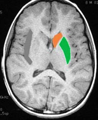
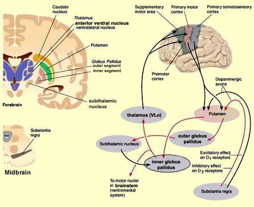
THE BASAL GANGLIA
The grouping formed by the caudate
nucleus (orange) and the putamen (green) is called the striatum.
It constitutes the major target for the cortical afferents of the basal ganglia.
The efferents from the basal ganglia to the thalamus arise in the globus pallidus.
The part of the ventrolateral nucleus of the thalamus that then projects to Area
6 is called "pars oralis" and usually designated by the symbol VLo.
The
other structures of the basal ganglia form various internal loops that modulate
the activity of the main loop, in which information passes through the following
brain structures in succession: cortex – striatum – globus pallidus
– VLo – cortex (supplementary motor area, or SMA).
We now know which of the connections in this main
loop are excitatory and which ones are inhibitory. They are illustrated in the
diagram below, as are the excitatory influence of the substantia nigra and the
subthalamic nucleus on various parts of this circuit.
Source: Jacob L. Driesen, Ph.D.
By
considering the interactions among the various structures in this loop, we can
get an overall understanding of how it operates. For example, we know that when
this loop is at rest, the neurons of the globus pallidus are spontaneously active
and consequently inhibit the VLo of the thalamus. But when this loop is activated
by a signal from the cortex, the neurons of the putamen are activated, thus inhibiting
those of the globus pallidus. Because the globus pallidus is suddenly less active,
its inhibitory effect on the VLo cells is removed. The resulting activation of
the VLo facilitates the activity of the SMA.
Thus we see that this is
a positive feedback loop that can focus information from wide areas of the cortex
onto the supplementary motor area. We can therefore posit that the signal that
ultimately triggers a voluntary movement occurs when the activation of the SMA
reaches a certain threshold under the influence of this loop.
Among the various learning-related
activities in which the cerebellum appears to be involved, one is the adaptation
of a certain number of reflexes, such as the vestibulo-ocular reflex.
This is the reflex that lets you keep looking in one direction while you turn
your head in another, by moving your eyes in the opposite direction. This reflex
can be modified with learning, and some injuries to the cerebellum can prevent
this learning.
Learning of conditioned reflexes also can be disturbed
by injuries to the cerebellum. One example is the palpebral reflex,
which makes you close your eyes automatically when a stream of air is directed
at them. This reflex can be conditioned by making subjects hear a certain sound
just before the stream of air is directed at their eyes. After several associations
of this kind, the sound alone will trigger the closing of the subjects' eyelids.
But if the cerebellum is damaged, this conditioned reflex cannot be learned, or
will be suppressed if it was learned previously.
The cerebellum 's circuits include
a system that can measure time, thus enabling the cerebellum to sequence various
functions that it controls. For example, someone with a damaged cerebellum will
have much more trouble in estimating the time interval between two sounds and
comparing them with a control interval.
Damage to this timing system
would explain why some people make mistakes when they use the information from
their sense of sight to calculated the speed of movement of their body parts or
other objects. It would also explain their poor motor coordination in both the
acceleration and the braking phases of body movements.
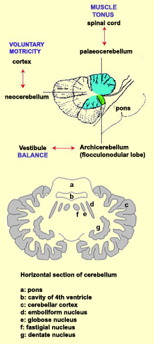
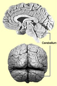
THE CEREBELLUM
The
pathologies of the cerebellum have long revealed that this part of the brain is
involved in motor co-ordination (see sidebar). The cerebellum is divided into
three regions, each of which is connected to a specific structure in the brain
and involved in a specific function.
The archicerebellum (or vestibulocerebellum)
first appeared in fish. It is connected to the vestibule of the inner
ear and is involved in balance.
The palaeocerebellum(or spinocerebellum) consists
mainly of the vermis, an axial structure, and is superimposed on the
archicerebellum in amphibians, reptiles, and birds. The palaeocerebellum is connected
to the spinal cord and controls postural muscle activity by influencing muscle
tonus. To play its role in maintaining body posture, a muscle must be tensed.
The cerebellum therefore controls muscle tension at all times while releasing
those muscles required to execute movements.
In mammals, the neocerebellum(or cerebrocerebellum)
is superimposed on these two other parts. It is more voluminous in primates and
especially so in humans. It consists of the cerebellar hemispheres, is connected
to the cortex, and contributes to the co-ordination of voluntary movements. Among
other functions, it ensures that when one set of muscles initiates a movement,
the opposing set acts as a brake, so that the body part in question arrives at
its target precisely.
The grey matter of the cerebellum is also organized
somewhat like the grey matter of the cerebral hemispheres: a cortex that forms
the grey matter at the surface, and deep nuclei that serve as relays for the efferent
pathways leaving this cortex . There are four of these cerebellar nuclei on either
side of the median line: the fastigial nuclei, also known as
the roof, serve as relays for the archicerebellum; the emboliform and
globose nuclei do so for the palaeocerebellum; and the dentate
nuclei, located in the middle of each cerebellar hemisphere, do so for
the neocerebellum.
For the body to make any
given gesture, the sequence and duration of each of the basic movements of each
body segment involved must be controlled in a very precise manner. One of the
cerebellum's jobs is to provide this control over the timing of the body's movements.
It does so by means of a loop
circuit that connects it to the motor cortex and modulates the signals that
the motor cortex sends to the motor neurons.
In humans, the cerebellum
also plays a role in analyzing the visual signals associated with movement. These
signals may come either from the movement of objects within the field of vision
or from the sight of the moving body segments themselves. The cerebellum appears
to calculate the speed of these movements and adjust the motor commands accordingly.
Errors in such calculations largely account for the poor motor control observed
in patients who have suffered injuries to the cerebellum.
As regards cognitive impairments, some signs of
cerebellar involvement have been found in the areas of language, attention, memory,
and emotions. For example, in some autistic children, cognitive delays have been
partly attributed to insufficient development of certain parts of the cerebellum.
Cerebellar syndrome
is the term used to designate manifestations of damage to the cerebellum, regardless
of origin (injury, tumour, stroke, etc.). For example, if a patient with cerebellar
syndrome tries to touch an object, the movement of his hand will begin late, then
accelerate beyond what is normal. Braking also will be too late, and inefficient,
so that his hand ends up missing the object and going past it. This movement then
ends with oscillations of the hand and arm.
People with cerebellar syndrome
also appear to have some problems in co-ordinating balance and posture. These
people have an uncertain gait, spreading their feet more widely apart as they
strike the ground. If these people are jostled, the reflexes that compensate for
the imbalance overreact, often resulting in oscillations of the entire body. These
people also cannot tilt their trunks forward or backward without losing their
balance.
The operation of each hierarchical
level in the motor control system is extremely dependent on the sensory information
that it receives. So much so, in fact, that to be fully understood, the motor
system must really be considered in sensorimotor terms. In the determining of
motor strategies, sensory information helps to generate a mental image of the
body and its position in its environment. The decisions on how to apply motor
controls (for example, the duration and amplitude of each contraction) are based
on memories of sensory information about past movements. And in the actual execution
of a movement as such, sensory feedback enables the brain to maintain the body's
posture and helps it to determine the length and tension of the muscles before
and after every voluntary movement.
THE ACTIVATION SEQUENCE FOR THE MOTOR AREAS
Any voluntary movement
can be accurately described as an intentional effort undertaken jointly by the
motor cortex and numerous other neural systems acting in a "consulting capacity".
This effort is organized hierarchically. First, the top level of the hierarchy
takes care of defining the motor strategies: the objectives of the movement
and the behaviours to be applied to achieve these objectives. When you decide
to take an elevator, for example, which will involve walking over to the Up button
and pressing it, your prefrontal cortex prepares the plans for this movement.
Meanwhile, your frontal cortex is receiving information from a large number of
axons projecting from the parietal cortex, which is involved in spatial perception.
Its analysis of the position of your body and its various members in space will
accordingly be essential to preparing for the movement. The
basal ganglia are another set of brain structures involved in this part of
the process.
Second,
the secondary
motor areas (PMA and SMA) work with the cerebellum
to specify the precise sequence of contractions of the various muscles
that will be required to carry out the selected motor action, in this case, raising
your arm and stretching your index finger out to the elevator button. But to do
this, your brain will need to convert the elevator button's location in the external
environment into a set of intrinsic co-ordinates that will let you adjust the
angles of the various joints that will be involved in the movement.
Third,
the primary
motor cortex, the brainstem, and the spinal cord come into play to produce
the contractions of all the muscles needed for the chosen movement.
The primary motor cortex determines how much force each muscle group must exert,
and then sends this information to the spinal motor
neurons and interneurons that generate the movement itself, as well as the
postural adjustments that accompany it.
For another example, here is how these three levels
work together when you throw a baseball. First, using the visual, auditory, somatic,
and proprioreceptive information provided by your sensory organs, your cerebral
cortex determines your body's position in space. The cortex exchanges information
with the basal ganglia about your goal in throwing the ball (for instance, whether
you want to throw it as high, as far, or as hard as possible) and the strategy
to adopt to achieve this goal, based on such things as your past experience in
throwing balls. Next, the secondary motor areas in your cerebral cortex and cerebellum
make the appropriate decisions concerning the amplitude, direction, and force
of the movements to make with your arm. These areas send these instructions to
your brainstem and cervical spinal cord, which trigger a co-ordinated movement
of your shoulder, elbow, wrist, and fingers. Simultaneously, commands sent to
the thoracic and lumbar spinal cord from the brainstem determine the postural
adjustments that will let you keep your balance while optimizing your movement
as you throw the ball. The motor neurons in your brainstem will also be activated
to keep your eye on the target that you are throwing at.
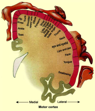 These
two elongated regions face each other, and the nerve fibres leaving and entering
them have the same somatotopic organization: they are like maps
that reproduce the anatomy of the human body on a small scale. But both in the
motor cortex and in the somatosensory cortex, the scale of this map is not constant.
In the motor cortex, it varies with the precision of the movements controlled
in the body part in question. In the somatosensory cortex, it varies with each
body part's sensitivity to sensory information.
These
two elongated regions face each other, and the nerve fibres leaving and entering
them have the same somatotopic organization: they are like maps
that reproduce the anatomy of the human body on a small scale. But both in the
motor cortex and in the somatosensory cortex, the scale of this map is not constant.
In the motor cortex, it varies with the precision of the movements controlled
in the body part in question. In the somatosensory cortex, it varies with each
body part's sensitivity to sensory information.