|
|

Through genetic manipulations
that enabled mouse embryo cells to produce melanopsin,
researchers have been able to make these cells photosensitive.
Likewise, other researchers have shown that frog eggs also
became light-sensitive when manipulated to express the gene
for melanopsin. Following a similar protocol, a third research
team has even succeeded in making an embryonic human kidney
cell light-sensitive . In fact, the cells in all of these
experiments began to behave like the subpopulation of retinal
ganglion cells that contain melanopsin, thus clearly indicating
that this pigment makes cells intrinsically photosensitive.

|
|
|
| LIGHT-SENSITIVE GANGLION CELLS |
|
Thanks to various tagging
methods, scientists now know that a certain sub-population
of the ganglion cells in the human retina contain a photosensitive
pigment and project
their axons directly into the suprachiasmatic nuclei as well
as into other brain structures that are concerned with
the intensity of ambient light.
These light-sensitive ganglion cells have large receptive fields, because of
their long,
widely dispersed dendrites. In these cells, accurate reception of information
on shape, orientation, and movement is sacrificed to general sensitivity. These
cells clearly constitute another light-sensitive system that runs parallel to
the visual system but is dedicated to detecting light intensity rather than to
forming images.
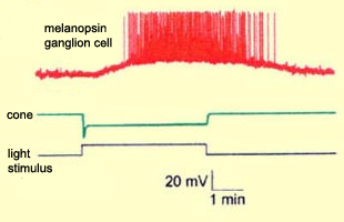
| The number of these light-sensitive
ganglion cells in each human retina is relatively small (only
about 2000). Their electrophysiological response to light stimuli
is quite different from that of the retina’s rods and
cones. In response to light, rod and cone cell membranes hyperpolarize
rapidly, but the membranes of these photosensitive ganglion
cells instead depolarize, and far more slowly. This type of
response is similar to that found in the photosensitive cells
of invertebrates such as flies and octopi, which supports the
idea that phylogenetically, this signalling system is far older
than the visual one. |
|
Because they respond to light stimuli so slowly, these ganglion
cells can integrate information over a long period—up to
5 minutes, according to some authors. This is exactly what you
would expect of a non-visual system dedicated to signalling the
overall intensity of light, rather than to transmitting
detailed information about visual images.
These ganglion cells, which receive their inputs from the amacrine
and bipolar cells in the retina’s inner plexiform layer, appear to
use two neurotransmitters: glutamate and pituitary adenylate cyclase-activating
polypeptide (PACAP).
Many experiments have shown that even though they do have indirect connections
with the retina’s better-known photoreceptor cells (the rod
and cones), these ganglion cells are also intrinsically photosensitive (see
sidebar). Their photosensitivity is, at
least in part, attributable to the fact that they contain the photosensitive
pigment melanopsin in their dendrites, their proximal axons, and their cell
membranes.
|
|