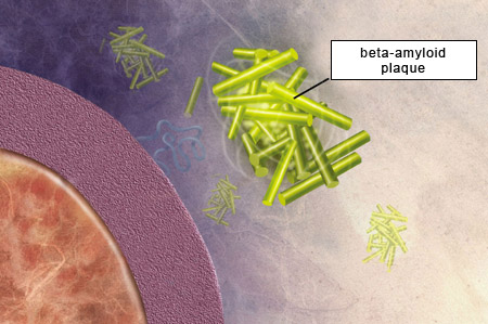
Funding for this site is provided by readers like you.
Les troubles de l'esprit
Sub-Topics

Depression and Manic Depression

Anxiety Disorders

Alzheimer’s- type Dementia


Linked
Help Link : The amyloid precursor protein: beyond amyloid
History
History : Alzheimer : historique des faits marquants
Original modules
History Module: Dr. Alois Alzheimer's First Cases Dr. Alois Alzheimer’s First Cases

Oligomers Help Us Keep Our Memories

Memory Loss in Alzheimer’s Reduced for the First Time

BETA-AMYLOID PROTEIN
THE TAU PROTEIN

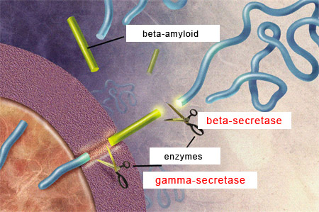
According to the amyloid hypothesis, the amyloid plaques that form between the brain’s neurons as it ages are toxic and hence cause the cognitive losses associated with Alzheimer’s. Although this hypothesis has not remotely explained all the mysteries of Alzheimer’s, researchers have devoted considerable effort to understanding the mechanisms by which these plaques form.

To begin with, what does the term “amyloid” mean? The answer goes back to the 19th century, when German physician Rudolf Virchow was studying a disease in which organs such as the heart, spleen, liver, and tongue swelled and hardened. To try to identify the substance responsible for this disorder, Virchow placed tissues from these organs in contact with Lugol’s solution, which contains iodine. According to the science of the day, if the tissues then changed colour, that would indicate the presence of glycogen or starch—in other words, chains of sugars.

Virchow found that Lugol’s solution did indeed cause the tissues to change colour. He attributed this change to a starch-like substance in the tissues, which he named “amyloid”, from the Greek amylo (starch) and -oid (similar to). But a few years later, it was shown that the substance that Virchow had stained was not a starch or sugar but a protein. Other stains, such as Congo red and thioflavin, also bound to this amyloid protein material. And in 1927, Belgian psychiatrist Paul Divry found that Congo red also stained the “senile plaques” that Alois Alzheimer had described two decades earlier.

For the next several decades, scientists tried in vain to determine exactly which protein formed the central component of what were now being called amyloid plaques. It was not until 1984 that American research pathologist George Glenner and his team characterized the structure of the protein molecules that agglutinate to form amyloid plaques. He discovered that what makes this protein “amyloid” is not the linear sequence of the amino acids that compose it, but rather its secondary structure—the way it is organized in space.

Certain proteins, or certain peptides (which are short proteins) fold in space into special structures known as beta-pleated sheets. And it is the high content of beta-pleated sheets in the protein (peptide) that Glenner was investigating that accounts for its affinity for Congo red and thioflavin—in short, for its amyloid nature. These beta-pleated sheets are also what makes this protein so compact, stable, and insoluble.

Because the peptide that Glenner identified had never been described previously, he named it A-beta peptide (A for amyloid and beta for the beta-pleated sheets). We now know that A-beta peptide, also commonly known as beta-amyloid, 
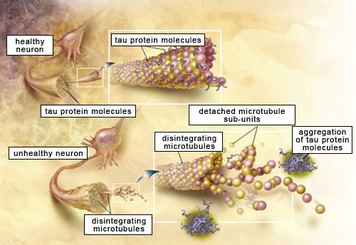
comes from the cleavage of  another, larger protein called amyloid protein precursor(APP).

APP is a transmembrane protein, which means that the APP molecule passes through the neuron’s cell membrane. APP is highly present in the central nervous system, where it is found at the synapses between neurons. APP, like the beta-amyloid that is derived from it, is thus a normal component of the organism. For example, APP helps neurons to grow, to survive, and to repair themselves when they suffer damage.

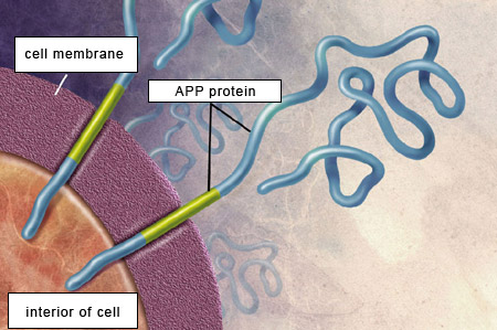
For the beta-amyloid peptide to be released, APP must be cleaved at two locations by two special types of enzymes. First, beta-secretases cut the APP’s chain of amino acids at a certain distance outside the cell membrane. Then gamma-secretases make another cut, this time inside the cell membrane, thus releasing the beta-amyloid peptide.

The length of this peptide varies from 38 to 42 amino acids. The two main forms of beta-amyloid have 40 and 42 amino acids, respectively, and the latter is the one that has the greater propensity to agglutinate and thereby form amyloid plaques.


Original modules
Experiment Module: The Effects of Normal Aging on Our Cognitive Abilities The Effects of Normal Aging on Our Cognitive Abilities

The importance of the role that the tau protein plays in the cognitive performance of older people is also supported by data obtained by less orthodox means. Most studies examine the brains of people displaying early memory loss and look for abnormalities that may explain this deficit. But in an article published in 2008, Changiz Geula and his colleagues reported that they had done the reverse: they had examined the brains of five very old people whose memories were exceptionally good for their age, and had looked for anything that might explain this fact.

All of these individuals were over 80 years old. Some of them even performed certain memorization tasks just as well as people in their 50s. After these subjects had died, Geula’s team compared their brains with those of people who had lived to the same age and had not developed dementia, but whose cognitive abilities had been more typical for their age.

Geula’s analyses showed that the brains of the elderly subjects who had excellent memories contained far fewer tangles of tau proteins than the brains of normal subjects. Also noteworthy was that the quantity of amyloid plaques was similar in the two groups, from which the researchers concluded that it was the smaller number of neurofibrillary tangles that seemed to be the key factor in elderly people with exceptional memories.

These individuals raise some doubts about the generally accepted idea that after age 75, tau pathologies are unavoidable. The subjects in this study seemed to have been largely spared any build-up of tau proteins, and the scarcity of neurofibrillary tangles in their brains is consistent with the memory performance that was so surprising in people of their age.

Link : “Super-aged” brains reveal secrets of sharp old-age memory
THE TAU PROTEIN
BETA-AMYLOID PROTEIN

In the 1980s, scientists were uncertain about the nature of the neurofibrillary tangles that are present in such large numbers in the neurons of people with Alzheimer’s. The prevailing hypothesis was that these tangles were composed of elements of the neuron’s cytoskeleton. In 1985, Belgian neuroscientist Jean-Pierre Brion found an antibody that selectively marks the neurofibrillary tangles in the brains of Alzheimer’s patients by binding not directly to the cytoskeletal proteins that give neurons their shape, but rather to the tau protein that stabilizes this cytoskeleton (tau stands for “tubulin associated unit”).

Other research teams then confirmed that the tau protein is the main component of neurofibrillary tangles. Like beta-amyloid peptide molecules, tau protein molecules are present in the body normally but become problematic when they undergo changes that increase their propensity to agglutinate (stick together). When they do so, they can no longer hold in place the microtubules—the “rails” that carry nutrients within the neurons—and thus compromise the neurons’ very survival.


Source: National Institute on Aging/U.S. National Institutes of Health


There are several suspected sources of the changes that might make tau proteins more likely to agglutinate: mutations in the gene for this protein, changes in the expression of this gene, anomalies during the steps leading to the production of the tau protein, etc.

But the number-one suspect appears to be excess phosphorylation of the tau protein. The tau protein can accept several phosphate groups at various sites on its molecular chain, and it may be that when too many of these sites are phosphorylated, the tau proteins become detached from the microtubules and start sticking to one another instead.

This process leading to the formation of neurofibrillary tangles is not specific to Alzheimer’s. It occurs in nearly 20 other dementias and neurodegenerative diseases, such as frontotemporal degeneration, Pick’s disease, and numerous Parkinsonian syndromes. The causes of tau-protein malfunction can vary for each of these conditions, but these changes always cause neurofibrillary tangles that ultimately impair cognitive functioning.

As a result, the concept of tau pathology or “tauopathies” has developed to describe the entire set of phenomena resulting from a direct or indirect malfunction of the tau proteins. Neuroscientists now believe that after age 75, this tau pathology appears even in the normal human brain (see the sidebar for a caveat, however), especially in the entorhinal cortex, and then in the hippocampus, two parts of the brain that are closely involved in long-term memory.

  Presentations | Credits | Contact | Copyleft