The term embryo refers
to the earliest stage of development of a human being, corresponding
roughly to the first two months of pregnancy. After that,
until the pregnancy ends, the future human being is called
a fetus. The fetus has all of the organs
of the human body, in rudimentary form.
From a medical standpoint, the duration of a pregnancy is calculated
from the first day of the last menstrual period. Actual fertilization
does not take place until 14 days later.
The first stage in the development of a human being, the embryonic stage, begins
with fertilization.
Fertilization generally occurs in the first third of the fallopian tube, the
canal that connects each ovary to the uterus. In fertilization, once one of the
spermatazoa (sperm cells) has penetrated the ovum, or oocyte,
it becomes impenetrable to all the other sperm cells.
Once fertilization has occurred, the primordial cell, called the zygote,
migrates to the lining of the uterus. While doing so, this cell
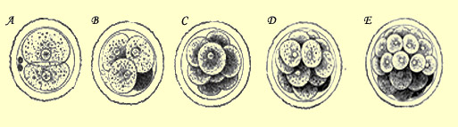
undergoes successive divisions, soon forming an embryo with 2 cells,
then 4, then 8, and so on.
a) 2-cell stage; b) 4-cell stage;
c) 8-cell stage; d) and e) morula stage
The first three-dimensional structure that emerges from these cell
divisions is a sphere of cells. The term morula is
used to designate the ensuing stages of embryonic development
(16, 32,and 64 cells). The morula is thus the product of the
first cell cleavages, which result in practically no growth,
because the daughter cells become smaller and smaller.
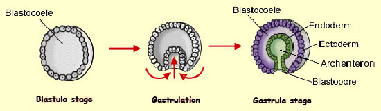
The morula is like a solid ball. But after the 64-cell stage,
this ball develops an inner cavity, called the blastocoele, thus
becoming a blastula. The blastocoele is bound
by a single layer of cells. It is during the blastula stage, about
7 to 8 days after fertilization, that the embryo becomes implanted
in the uterine wall.
Some cells of the blastula soon being moving toward the interior
of the blastocoele to form distinct layers that will be redistributed
as the blastula continues to invaginate (fold inward) in the next
developmental stage, known as gastrulation.
The blastula becomes the gastrula when the invaginated cells have
formed the ectoderm
and the endoderm.
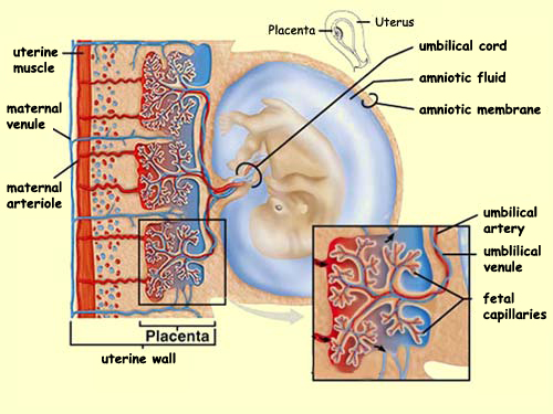
In pregnancy, the placenta develops
from the membrane surrounding the fetus and the uterine lining.
Attached to the wall of the uterus, the placenta is like
a spongy cake that supplies the fetus with nutrients and
oxygen through the umbilical cord. The placenta also enables
the fetus to eliminate its metabolic waste products and pass
then out into its mother’s bloodstream.
In addition, the placenta secretes a number of hormones, including
progesterone, estrogens, lactation-promoting hormones, and
a hormone called chorionic gonadotropin that is found in the
urine of pregnant women and that is the basis for pregnancy
tests.
But even as it enables all these intimate exchanges between
the mother and the fetus, the placenta also prevents their
blood from mixing. The placenta thus acts like a sort of border
patrol, preventing most types of germs from crossing over from
the mother to the fetus. But the mother’s antibodies,
and any drugs that she may take during pregnancy, do cross
the placenta. If the drug is a medication such as an antibiotic,
it may be helpful, protecting the fetus from infection. But
if the drug is alcohol or
some kind of street drug, it could have negative effects on
the baby’s development.
The closing of the
neural tube is a crucial event in the development of the
nervous system. This event in turn depends on a sequence
of events that affect the position of the cells and the
processes of adhesion between them. When the neural tube
fails to close correctly, serious birth defects can result.
One of the best known of these is spina bifida, which
occurs in about 1 of every 1000 births. It is caused by a
malformation of the caudal portion of the neural tube. This
malformation in turn results in a malformation of the lower
vertebrae that often leaves the spinal cord exposed, makes
it vulnerable to injury, and limits use of the legs and feet.
Spina bifida appears to be associated with a deficiency of
folic acid. This vitamin should be available in sufficient
quantities in the pregnant mother’s food, but if her
diet is poor or imbalanced, the resulting shortage of folic
acid can be serious enough to interfere with the formation
of the neural tube.
In the opposite condition from spina bifida, the upper portion
of the neural tube remains open. The result is a birth defect
called anencephaly, which is quite serious too. In
this condition, the
organization of the major structures of the brain is
greatly disturbed.
HOW THE NERVOUS SYSTEM BEGINS
Just as studying the evolutionary
origins of the human brain can teach us much about its anatomy,
studying how the nervous
system develops over an individual’s lifetime gives
us a better understanding of how this system is organized.
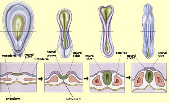
The formation of the nervous system occurs fairly early in embryonic development
and is referred to as neurulation. An important structure that appears at the
end of the preceding stage (the gastrulation stage)
is the dorsal cord, or notochord. This cylinder of cells in the mesoderm defines
the embryo’s rostral-caudal axis and extends along its
entire length.
Around the third week of gestation, the notochord sends a
molecular signal that causes the cells of the ectoderm just
above it to thicken into an individualized epithelial column,
the neural plate. After this “neural induction”,
the neural plate begins to invaginate to form the neural groove, which then rises
from the embryo’s surface and closes to form the neural
tube.
On the dorsal side of the neural tube, another special population
of cells is distinguished where the neural tube protrudes, whence
its name, the neural crest. These cells will eventually migrate
along specific pathways that will expose them once again to
various inductive
molecules. Ultimately, these cells will differentiate to form
structures such as the spinal and vegetative ganglia.
On either side of the neural tube, the mesoderm thickens and divides
into structures called somites. These are the
precursors of the axial musculature and the skeleton. The part
of the neural tube in the vicinity of the somites will form the
future spinal cord. The rostral end of the neural tube will close
and continue to grow to form
the various structures of the brain.
The body’s first movements begin
during fetal life. They consist essentially of reflex movements
such as sucking and grasping and spontaneous movements
such as stretching. The reason is that the first parts
of the brain to become functional are mainly subcortical
structures. These structures developed
earlier in the course of evolution and
are responsible for stereotyped movements such as the reflexes.
HOW
THE MAJOR SUBDIVISIONS OF THE BRAIN ARE FORMED
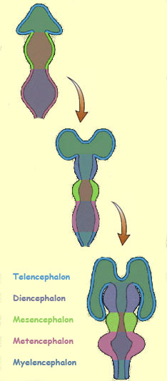
The stage in which
the more elaborate structures of the brain develop from
the neural tube is called differentiation.
The first structure to appear is an embryonic
brain composed of three primary vesicles. During the seventh week of development,
two of these vesicles themselves divide in two, so that there are then a total
of five secondary vesicles.
The rostral
part of the prosencephalon produces two lateral buds that
grow into the telencephalon—two large
vesicles that will ultimately become the cerebral hemispheres.
The posterior part of the prosencephalon forms the diencephalon, which
will comprise the thalamus, hypothalamus, pituitary gland,
pineal gland, and retina.
The middle member of the three primary vesicles, the mesencephalon,
does not subdivide. It evolves more slowly and ultimately
forms such structures as the tegmentum and the superior
and inferior colliculi.
The most caudal of the three primary vesicles, the rhombencephalon,
elongates rapidly. As a result, it must bend ventrally,
forming the pontine flexure. This flexure
divides the rhombencephalon into a rostral portion, the metencephalon,
which will become the pons and cerebellum,
and a caudal portion called the myencephalon,
which will become the medulla oblongata.