Funding for this site is provided by readers like you.
L'émergence de la conscience
Sub-Topics
The Sense of Self

Linked
HelpLien : La cartographie du système cérébralLien : Cortical networks for working memory and executive functions sustain the conscious resting state in manLien : A relation between rest and the self in the brain?
Lien : Functional connectivity in the resting brain: A network analysis of the default mode hypothesisLien : Tutorial commentary: surprisingly small subcortical structures are needed for the state of waking consciousness, while cortical projection areas seem to provide perceptual contents of consciousnessLien : Brain stem may be key to consciousnessLien : Pre-frontal executive committee for perception, working memory, attention, long-term memory, motor control, and thinking: A tutorial review
Lien : The central role of the parietal lobes in consciousnessLien : On the unity of conscious experienceLien : Definition of Centromedian nucleusLien : The Electric Brain
Lien : Reflective Self-Awareness and Conscious States: PET Evidence for a Common Midline Parietofrontal CoreLien : Neutraliser une région du cerveau pourrait combattre la dépendanceLien : L’empathie ou l’émotion partagéeLien : The precuneus: a review of its functional anatomy and behavioural correlates
Lien : Experiencing Oneself vs Another Person as Being the Cause of an Action: The Neural Correlates of the Experience of AgencyLien : Insular cortexLien : A Small Part of the Brain, and Its Profound EffectsLien : Interrupting the “stream of consciousness”: An fMRI investigation
Lien : Need To Rethink Thought: Human consciousness may be only 2,000 years oldLien : Are You Conscious of Your Precuneus?Lien : Science and Consciousness ReviewLien : La conscience emmurée
Experiment
Expérience : Neuroimaging of genesis and satiation of thirst and an interoceptor-driven theory of origins of primary consciousnessExpérience : Quand le cerveau bascule vers la perception conscienteExpérience : Clinicometabolic dissociation of cognitive functions and social behavior in frontal lobe lesions

Thalamocortical interactions are involved in generating neuronal oscillations across various parts of the cortex. In this sense, the thalamus acts somewhat like the conductor of an orchestra whose musicians are distributed throughout the cortex. The conductor doesn’t play the music for the musicians, but instead co-ordinates them and imposes a rhythm on them. Without the thalamus, the cortex could probably display some isolated examples of synchrony but would not be able to bind the various properties of a perception into a coherent concept .

This metaphor also helps us to understand why it is pointless to look for any one seat or centre of consciousness in the brain. The thalamic conductor might impose the rhythm, but that would be meaningless unless the cortical musicians were playing their sensory scores. It is the co-ordination of all these things that makes the mental symphony - the object of consciousness - coherent.


The thalamus is very well positioned to control the inputs to the cortex. Among the various thalamic nuclei, the reticular nucleus is known to exert an inhibiting modulation on the other specific sensory nuclei of the thalamus. The reticular nucleus thus helps to select the sensory inputs that can reach the cortex, and hence enter consciousness.

The circuits in this thalamic nucleus can favour one particular input over several others. For example, this is what happens when a stimulus that has a strong meaning for someone (such as the sound of their own name) manages to clear a path through numerous other auditory stimuli and thus reach that person’s consciousness.

This kind of activation, “from the bottom up”, would be controlled by the brainstem, the amygdala, or the systems associated with the perception of pain. In contrast, activation “from the top down” would be controlled by the executive functions of the frontal cortex and, according to certain authors, would operate through the anterior cingulate cortex.


Brodmann area 46, located in the frontal cortex, is activated by a wide range of tasks and seems well situated for co-ordinating conscious thoughts. In conjunction with all the other areas of the brain, area 46 might help us to switch from one thought to another by facilitating certain global activation patterns at the expense of others.

The particular content of a thought—“what you have on your mind”— is associated with the content of working memory: the temporary memory that you use for tasks such as doing arithmetic in your head, or keeping your train of thought while formulating long sentences or advancing complex arguments, or for assessing possible moves when you are playing chess.

This working memory is often described as being composed of a central executive (identified with frontal area 46) and two main auxiliary “slave” systems.

One of these auxiliary systems is a visual/spatial form of memory that engages several areas in the right hemisphere. This memory holds mental images, the pictures that you can “see in your head” and that are so helpful for solving spatial-configuration problems.

The second auxiliary memory system comprises an auditory form of memory, or “phonological loop”. This is the locus of your inner discourse, that small voice that you constantly use to talk to yourself and that activates the areas in the left hemisphere that are used to decode language.

But regardless of which of these two auxiliary systems is at work, the executive processor in the frontal lobes is always activated.

Lien : Brodmann area 46Outil : Les aires corticales de Brodmann

Embodied Cognition and Emotions

The Brain’s Default Network


To try to explain the complex role of the frontal cortex more clearly, some authors use the metaphor of an executive committee composed of five members, each of whom represents a sub-committee of more posterior or subcortical areas in the brain.

The first member is the Perceiver. Located mainly in the ventral-lateral portion of the right frontal hemisphere, the Perceiver is the frontal extension of the ventral perceptual system and is focused on objects. The second member is the Verbalizer. Dominant in the ventral-lateral portion of the left prefrontal cortex, it is the frontal extension of the language circuits. The third committee member is the Motivator. Located in the ventral-medial region of the orbitofrontal cortex, it is the cortical extension of subcortical pathways that include the amygdala and represent the world in terms of emotional motivations. The fourth member, the Attender, occupies the dorsal-medial portion of the frontal cortex, as well as the anterior cingulate cortex. The Attender is the frontal extension of a subcortical pathway involving the hippocampus. The Attender represents the world and self in spatiotemporal co-ordinates and can direct attention to internal and external events. Lastly, the fifth member is the Co-ordinator (or central executive - see preceding sidebar). It is located in the dorsolateral region of the frontal cortex and is the frontal extension of the dorsal pathway. The Co-ordinator represents the world and self in body-centred coordinates, which enables it to control willed movements and working memory.

Lien : Pre-frontal executive committee for perception, working memory, attention, long-term memory, motor control, and thinking: a tutorial review
CAN STATES OF CONSCIOUSNESS BE MAPPED IN THE BRAIN?

Some neurobiological models of consciousness, such as the global workspace theory, assume that the contents of consciousness are widely distributed in the brain. This assumption has been confirmed by many brain-imaging experiments, in particular those of Stanislas Dehaene and his collaborators. In these experiments, when the amount of time that a word was projected onto a screen was extended just past the threshold required for subjects to perceive it consciously, there was a major increase in activity in their frontal, prefrontal, anterior cingulate, and parietal cortexes.

Thus conscious sensory inputs would appear to produce far more extensive brain activity than comparable unconscious stimuli, and a sudden activation of the frontal and parietal lobes would appear to be the typical signature of a conscious perception.

But this perceptual consciousness, or as some would call it, primary consciousness, is not the only form of consciousness. When we are trying to associate consciousness with particular structures in the brain, we must therefore clearly define what level of consciousness we are talking about. For example, the first condition necessary for the brain to be able to process external sensory stimuli consciously is that it must be in an appropriate state of alertness (for instance, awake rather than asleep).

Starting from this premise, authors such as Damasio distinguish a very primitive form of consciousness that he calls the proto-self and that is more like a moment-to-moment perception of the body’s internal emotional state. This state is associated with activity of such brain structures as the reticular formation, the hypothalamus, and the somatosensory cortex.

The reticular formation is also associated with consciousness in the minimal sense of wakefulness. Other structures involved in simply maintaining wakefulness include the pons, the raphe nuclei and the locus coeruleus.

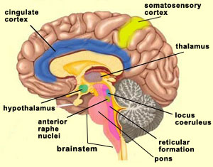
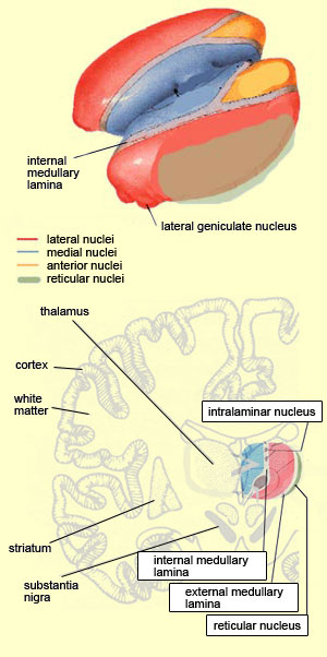
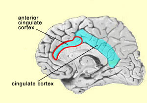
It should be noted here that the activity of the reticular formation, like that of the primary sensory areas, seems to be necessary but not sufficient for a more elaborate level of consciousness. This latter level is attained with what several authors call primary consciousness, meaning a waking state in which we are in relationship with our environment “here and now”. On the basis of the research done by Swedish neuroscientist Bjorn Merker, it seems that the brainstem plays a more important role in primary consciousness than was formerly believed.

Damasio calls this type of consciousness “core consciousness” and says that it depends chiefly on the cingulate cortex and on the intralaminar nuclei of the thalamus. Indeed, experiments have shown that bilateral destruction of the centromedial portion of the intralaminar nuclei of the thalamus also eliminates consciousness, produces a coma, or causes other states similar to brain death. In addition, this region of the thalamus is one of the main sites acted upon by anaesthetics and by antipsychotic drugs.

Models of consciousness that attribute a role to the thalamus are no recent development. As far back as 1984, Francis Crick offered one of the first hypotheses about consciousness, the “thalamic searchlight hypothesis”, according to which the thalamus controlled which region of the cortex became the focal point for consciousness. A similar but more sophisticated idea has recently been proposed by Rodolfo Llinas. He hypothesizes that the oscillations of certain neurons in the thalamus serve as a sort of basic rhythm with which the cortical oscillations of the various sensory modalities synchronize themselves to form a unified image of the environment—somewhat like an orchestra conductor who provides the beat for all the musicians to follow (see sidebar). This is an original solution to the binding problem.


Lien : Thalamus Lien : Thalamus humain

The thalamus is often compared to a railroad switching yard, because the signals from all of the senses (except smell) must pass through it before they can reach the cortex. The cortex also sends many connections back to the thalamus. Most of the nuclei in the thalamus are considered “specific” because their neurons make connections with relatively circumscribed areas in the cortex (for example, the neurons of the lateral geniculate nucleus project to the primary visual cortex).

The thalamus also has many “non-specific” nuclei that send diffuse projections to wide areas of the cortex. The intralaminar nuclei, located in the internal medullary lamina, are a good example of non-specific thalamic nuclei.

To complete this overview of the thalamus, we should note that only one of its nuclei, the reticular nucleus, which wraps around the lateral portion of the thalamus, does not send any projections directly to the cortex. It does, however, play a role in the thalamocortical feedback loops, by receiving inputs from the cortex and sending outputs to the dorsal nucleus of the thalamus.


These thalamocortical loops have come to play an important role in practically all of the neurobiological theories that attempt to explain the higher states of consciousness, for which the lower levels of consciousness that we have been discussing up to now are in a sense only the prerequisites. These higher levels of human consciousness are known as reflexive consciousness and self-consciousness.

Reflexive consciousness—this sense that “it is I who am perceiving”—is often presented as a necessary condition for self-consciousness: the feeling of being oneself and not someone else. This autobiographic dimension of consciousness implies that we can form mental representations of conscious experiences in the past or the future, and therefore requires the support of memory and the higher functions that make abstract conceptualization and planning possible.

You would therefore expect that the areas of the brain that are known to be involved in these functions, especially in the frontal and parietal lobes, would be actively engaged in self-consciousness. And that indeed has been found to be so in certain studies that addressed this specific question.

These higher levels of consciousness also appear to involve other brain structures whose roles were long poorly understood, partly because some of them are located deep in the brain, which made them hard to access. Modern brain imaging techniques have now overcome this problem.

Three of these structures—the angular gyrus, the precuneus, and the anterior cingulate cortex, which are often very active in a conscious state of rest—may be part of a functional network that makes self-consciousness possible.

The case of the precuneus, which is the postero-medial portion of the parietal lobe, is especially revealing. The conscious resting state is a state in which the subject’s eyes are generally closed and the subject’s EEG shows an alpha rhythm, or in which the subject is passively looking at a simple target such as a “+” sign. Among all the areas of the brain that are active during this state, the precuneus is the one that shows the highest rate of neural activity. But in contrast, the precuneus is known to be less active during tasks that make no reference to the self. Some authors have therefore proposed that the activation of the precuneus, and of the posterior cingulate cortex, which is closely connected to it, is correlated with the feeling of selfhood and the sense of being an “agent”.


After Wheatley et al., 2007.

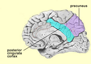
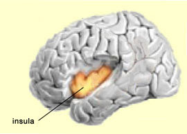
This hypothesis is also consistent with studies that have shown decreased activity in the postero-medial parietal cortex in many states of altered consciousness, such as sleep, anaesthesia, or a vegetative state. Other studies have also shown decreased activity in the precuneus and the posterior cingulate cortex when the subject is under hypnosis, another altered state of consciousness.

Lastly, the precuneus also seems to play a role in visual/spatial imagery. For example, some experiments have shown that the precuneus is more active when subjects are is engaged in motor imagery of a finger movement than when they are actually performing this movement. This again seems to indicate that people have a propensity to represent their own bodies in space.

The insula (also known as the insular cortex) is another region of the brain that remained little understood for a long time because of its position deep in the folds of the cortex. Also, because it was not associated with the “higher” brain functions, it was of less interest to scientists who were investigating consciousness.

But this indifference gave way to intense interest after Antonio Damasio conducted research on the insula and proposed that most of this structure consists of somatic markers. Damasio hypothesized that this part of the cortex maps the bodily states associated with our emotional experiences, thus giving rise to conscious feelings. This hypothesis falls within the school of thought known as embodied cognition, according to which conscious rational thought cannot be separated from emotions and their incarnation in the rest of the body.


After Wheatley et al., 2007.

The insula thus appears to provide an emotional context that is suitable for a given sensory experience. The insula is also well positioned to integrate information about the state of the body and to make this information available to higher-order cognitive and emotional processes. For example, the insula receives homeostatic sensory inputs via the thalamus and sends outputs to several structures associated with the limbic system, such as the amygdala, the ventral striatum, and the orbitofrontal cortex.

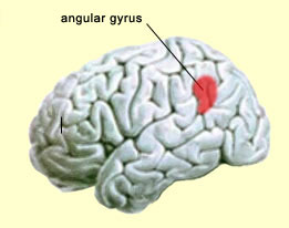
The insula has also been convincingly shown to be associated with pain processes, as well as with several basic emotions such as anger, fear, disgust, joy, and sadness. Its most anterior portion is regarded as part of the limbic system. The insula also appears to be deeply involved in conscious desires, such as the active search for food or drugs. What is common to all of these states is that they affect the entire body profoundly—which tends to strengthen the case for the insula’s likely role in the way we represent our bodies to ourselves and in the subjective aspect of emotional experience.

Lastly, the insula in humans, and to a lesser extent in the great apes, appears to incorporate two evolutionary innovations that provide these species with a greater ability to read the state of their own bodies than any other mammals.

First, the anterior portion of the insula, and more particularly that of the insula in the right hemisphere, is more developed in humans and great apes than in other animal species. This greater development might enable more precise decoding of bodily states—the capability that translates a bad odour, for example, into a feeling of disgust, or the touch of a lover into a feeling of delight.

The other major evolutionary modification in the insula is a type of neuron that is found only in the great apes and in humans. These large, elongated, cigar-shaped nerve cells are known as von Economo neurons (VENs). VENs occur only in the insula and in the anterior cingulate cortex. These neurons connect to various parts of the brain, which would be an essential attribute for the higher functions attributed to these two brain structures.

Now it is time to say a few words about the anterior cingulate cortex, which also acts as an important interface between emotion and cognition, and more specifically in the conversion of feelings into intentions and actions. This structure is involved in higher functions such as controlling one’s own emotions, concentrating on solving problems, recognizing one’s own mistakes, and making adaptive responses to changing conditions. All of these functions are closely linked with our emotions.


After Wheatley et al., 2007.

When experimental subjects are pricked with a needle, the activity in their cingulate cortex increases; this response is so clear-cut that the neurons in question are often called the “pain neurons”. A fascinating side-note: in 1999, William Hutchison and his colleagues at the University of Toronto showed that these same neurons in the cingulate cortex also become active when the subject sees someone else being pricked with a needle. Thus, for these kinds of neurons, known as mirror neurons, there is no boundary between the self and the other.

Primates, including humans, are highly social creatures. Knowing other individuals’ intentions has always been crucial for our survival. That is why we are past masters of the art of internally simulating other people’s minds, perhaps with the help of such mirror neurons.

Some neuroscientists, such as V.S. Ramachandran, even suggest that this ability to decode other individuals’ states of mind may even have evolved first, and subsequently been applied to the self, to become what we call self-consciousness. And in Ramachandran’s view, not only the mirror neurons, but all parts of the brain that contribute to language, such as Wernicke’s area in the temporal lobe, must inevitably play a role in this process.

This important role is ascribed to language in several models of higher consciousness, including that proposed by Michael Gazzaniga, who is known for his work with “split-brain”patients. But while Gazzaniga's model identifies the language hemisphere as the locus of this “interpreter” that makes us conscious of ourselves, other authors, such as Edelman, argue that consciousness cannot be attributed to any specific structure in the brain.

  Presentations | Credits | Contact | Copyleft