|
|


|
|
| AMYLOID PLAQUES AND NEUROFIBRILLARY TANGLES |
|
Amyloid plaques and neurofibrillary tangles are the two main biological markers associated with Alzheimer’s.
Dr. Alois Alzheimer himself observed them in a female patient, Auguste D., in the very early 20th century (follow the History Module link to the left). But even now, much about them remains unknown. As one brain scientist put it ironically, “At this point, we know almost everything about Alzheimer’s,
except for the role of amyloid plaques and neurofibrillary tangles.”
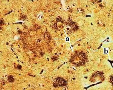
And yet these two types of lesions have been observable under the optical microscope since Dr. Alzheimer’s time, by a method called silver staining. When silver stain is applied to brain tissue, it causes silver salts to precipitate, somewhat like when a photograph is developed. These salts precipitate preferentially onto fibrillary cell structures, such as amyloid plaques and neurofibrillary tangles, thus making them visible under the optical microscope, as in the photo below.
Silver-stained cross-section of human cortex, showing several amyloid plaques (a) and blood vessels (b)
|
In what is considered their “classic", focal form, amyloid
plaques are spherical fibrous masses 30 to 100 micrometres in diameter that build up outside the neurons (in between them). The core of these plaques consists of a deposit of molecules of the peptide beta-amyloid, which results from the cleavage of a transmembrane precursor protein, known as amyloid
protein precursor, or APP. Beta-amyloid,
like APP, is a substance normally found in the body, though its function is still poorly understood. |
In the classic form of amyloid plaques, extensions of the nerve cells (mainly axons) wrap around the outside of the inert core. But plaques can take other forms as well; sometimes the amyloid deposits are more diffuse, with ill-defined borders and no peripheral neural extensions.
The amyloid cores of plaques also contain microglial cells, the brain’s scavenger cells, which cause a moderate inflammatory reaction there.
Neurofibrillary tangles are found inside neurons and are composed of a substance whose molecules clump together to form fibres with a very small diameter, on the order of 10 nanometers. Neurofibrillary tangles were first observed in 1961, under the electron microscope, by Michael Kidd and his team. At first Kidd and his colleagues thought that these structures were normal but modified components of the neurons’ microtubules, but then the researchers showed that these were abnormal structures, composed of paired helical filaments (PHFs).
PHFs themselves are composed of molecules of tau protein ("tau” stands for “tubulin associated unit"). Normally, these molecules are positioned perpendicularly to the microtubules, keeping them stable so that they can transport nutrients and other molecules within the cells and, in the case of neurons, along the axons.
Under normal conditions, some tau protein molecules become detached from the microtubules from time to time, but break down rapidly and are replaced. However, when certain chemical changes occur—specifically, when too many phosphate groups are added to the tau protein molecules—they can become “sticky”. As a result, they wind around one another to form paired helical filaments.
As these PHFs build up in the neuron’s cell body and its extensions, they destabilize the microtubules, compress and damage the neuron, and eventually cause it to die. |
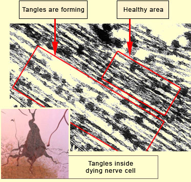
This photo taken with an electron microscope show a cell that has some healthy areas and other areas where tangles are forming.
Source: Alzheimer Society Brain Tour: More About Tangles |
This neuronal death causes chemical imbalances in neuronal communication processes and, years later, the first signs of cognitive deficits, in particular memory loss.
These two degenerative processes—the formation of amyloid plaques and neurofibrillary tangles—are common in the course of normal aging as well,
and have been fairly well described individually. What is less clear is the relative importance of each of them in the development of Alzheimer’s disease.
A number of biochemical and neuropathological findings suggest that the two processes tend to potentiate each other.
In the most sophisticated animal models, researchers have included some mutations in the two proteins in an attempt to reconstruct the two types of lesions and model their synergy.
Many members of the scientific community see this synergy as asymmetrical.
Most researchers believe that Alzheimer’s is expressed first following the buildup of amyloid plaques. But some researchers think that it is actually expressed with the appearance of neurofibrillary tangles. There are many other promising avenues for research that have yet to be explored, because the amyloid hypothesis on the origin of Alzheimer’s has been so predominant, but that is starting to change.
|
|