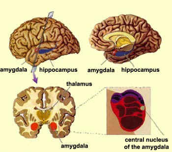
We now know that the brain comprises several different kinds of memory. The hippocampus and the cortex make explicit, conscious memories possible. For its part, the amygdala enables one of the forms of implicit memory: emotional memories associated with fear.
Various aspects of an especially emotional situation such as a car accident will therefore be processed both by the hippocampus and by the amygdala, working in parallel. Thanks to the hippocampus, you will remember whom you were with, what you did, and the fact that it was a particularly painful situation. However, it is because of the amygdala that when you remember the event, your palms will sweat, your heart will race, and your muscles will tense.
Suppose you are walking down the street when an unsavoury-looking character suddenly assaults you. A few days later, someone starts running toward you, and your heart begins to pound. The person runs past you without touching you, and you start to calm down. It turns out they were just running to catch a bus.
A few weeks after that, you pass by the place where you actually were attacked, and you feel sick. This time no one is running toward you. The conditioned stimulus is not present, but the situation reveals a common phenomenon, in which certain elements of the context have also been conditioned by the traumatic event. This phenomenon implies the involvement of the hippocampus.
THE AMYGDALA AND ITS ALLIES
The
amygdala is a brain structure that is essential for decoding
emotions, and in particular stimuli that are threatening
to the organism. As a result of evolution, many
of our body’s alarm circuits are grouped together in
the amygdala.
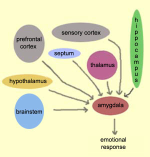
But there are several other regions of the brain that project
their axons to the amygdala; examples include the hypothalamus,
the septum and the reticular formation of the brainstem.
The hippocampus also specializes
in processing sets of stimuli (as opposed to individual
stimuli)–in other words, the context of a situation.
Hence it is because of the hippocampus and its close connections
with the amygdala that the entire context associated with
a traumatic event can provoke anxiety.
Major connections to the the amygdala also come from the medial prefrontal
cortex. These connections appear to be involved in the process
of extinction, whereby a stimulus that triggers a conditioned
fear gradually loses this effect. This happens if that stimulus
is repeatedly presented to the subject without the unconditional
nociceptive stimulus that was initially associated with it to
produce the conditioned fear.
The prefrontal cortex also seems to
be involved in the final phase of confronting a danger, where,
after the initial automatic, emotional reaction, we are forced
to react and choose the course of action that can best get us
out of danger. In people whose frontal cortex is damaged (people
with “frontal syndrome"), planning
the slightest task is very difficult, if not impossible.
Thus, the ability that our superior mental structures give us
to voluntarily plan an emotional response suited to the situation
is a wonderful complement to our
system of rapid, automatic responses. The connections from
the prefrontal cortex to the amygdala also enable us to exercise
a certain conscious control over our anxiety. However, at the same
time, this faculty can create anxiety by
allowing us to imagine the failure of a given scenario or even
the presence of dangers that do not actually exist.
The “wiring” of the body’s
natural alarm system demonstrates the usefulness, from an
evolutionary standpoint, of the automatic reactions evoked
by fear. For example, a small rodent that sees a predator
will freeze in place automatically. This automatic reaction
is invaluable, because it happens fast, with no need for
voluntary control on the rodent’s part. The rodent’s
immobility, combined with its natural camouflage, will generally
let it escape the predator’s notice and flee as soon
as its back is turned. As mammals evolved, those rodents
that were less “fearful” and did not freeze in
place attracted predators’ attention more quickly and
thus, though very courageous, did not leave many descendants.
If researchers condition a rat to
fear a certain sound, and then surgically remove the rat’s
auditory cortex, the rat will no longer be able to distinguish
that sound. A human with equivalent damage would be considered
deaf. Yet the rat, once recovered from its operation and
to all appearances deaf, still shows fear reactions when
the sound is made in its presence. The rat thus still seems
to register the sound in its thalamus and amygdala, which
suffices to trigger the fear reaction.
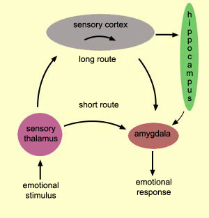
THE TWO PATHWAYS OF FEAR
Information from an external stimulus reaches the amygdala
in two different ways: by a short, fast, but imprecise route, directly
from the thalamus; and by a long, slow, but precise route,
by way of the cortex.
It is the short, more direct route that lets us start preparing
for a potential danger before we even know exactly what it
is. In some situations, these precious fractions of a second
can mean the difference between life and death.
Here is an example.
Suppose you are walking through a forest when you suddenly
see a long, narrow shape coiled up at your feet. This snake-like
shape very quickly, via the short route, sets in motion
the physiological reactions of fear that are so useful
for mobilizing you to face the danger. But this same visual
stimulus, after passing through the thalamus, will also
be relayed to your cortex. A few fractions of a second
later, the cortex, thanks to its discriminatory faculty,
will realize that the shape you thought was a snake was
really just a discarded piece of garden hose. Your heart
will then stop racing, and you will just have had a moment’s
scare.
But if your cortex had confirmed that the shape really was
a snake, you probably would not have just been startled. You
would probably have taken off with all the alacrity that the
physiological changes triggered by your amygdala allowed.
Thus, the fast route from the thalamus to the amygdala does
not take any chances. It alerts you to anything that seems
to represent a danger. The cortex then makes appropriate adjustments,
suppressing any reactions that turn out to be inappropriate.
Thus, we see, from an evolutionary perspective, how these two
complementary pathways may have become established. From the
standpoint of survival, the consequences of mistaking a garden
hose for a snake are less severe than those of mistaking a
snake for a garden hose.
But the cortex is not the only part of
the brain that puts in its two cents by specifying the nature
of the object. The hippocampus can also come into play by giving
you information about context.
In
the conditioned-fear protocol, an animal can be taught
to be afraid when it hears one particular sound, but
not when it hears another sound that is only slightly
different. But if you destroy this animal’s auditory
cortex, then it will be just as afraid of the sound
that is slightly different!
The
reason has been discovered in electrophysiological
experiments in which the activity of neurons in the
thalamus and the cortex was recorded. The cortical
neurons responded to only a very narrow range of
frequencies, while the thalamic neurons were activated
in response to a very broad range.
Consequently,
when two similar sounds are used to condition a fear,
and the cortex is removed to eliminate the possibility
of fine discrimination, the fear response that is controlled
by the thalamus will be displayed for both auditory
stimuli without distinction.