|
|
|
|
 |
| Fear,
Anxiety and Anguish |
|
|
|
|


Lesions to the central
nucleus of the amygdala interfere with just about all of
the manifestations of conditioned fear, including behavioural
inhibition, autonomic
nervous system responses, the suppression of pain, the
release of stress hormones, and the potentiation of reflexes.
In addition, each of these responses
is controlled by various bundles of nerve fibres projecting
from the central nucleus. For example, the nerve fibres projecting
to the periaqueductal grey matter interfere with behavioural
inhibition, but not with blood pressure. Conversely, the
fibres projecting to the lateral hypothalamus prevent blood
pressure from increasing, but do not affect behavioural inhibition.
Meanwhile, a lesion to the projection to the bed nucleus
of the stria terminalis has no effect on blood pressure or
behavioural inhibition, but does interfere with the release
of stress hormones. |
|
|
| THE CIRCUITS OF
THE BODY'S ALARM SYSTEM |
|
Because neuroanatomists distinguish
many different nuclei within the amygdala,
the term “amygdaloid complex” is often preferred
to denote this structure within the brain. This term is all the
more appropriate in that some of these nuclei can be further
broken down into various divisions. The following table lists
these nuclei and their divisions.
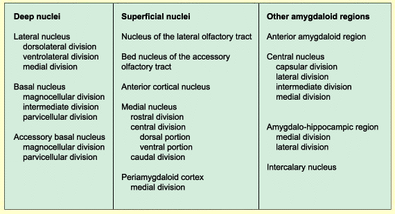
Source: Asla Pitkänen, Vesa Savander
and Joseph E. LeDoux Trends Neurosci. (1997) 20, 517-523
Given all these nuclei and their many divisions,
how does information circulate within the amygdaloid complex? Many
studies have helped to reveal the wiring that enables the amygdala
to detect potentially dangerous stimuli and orchestrate an appropriate
physiological response.
First it was noted that the projections from
the sensory regions of the brain enter the amygdala via the lateral
nucleus, which constitutes the main gateway to the amygdala, though
not the only one.
Some projections from various parts of the
brain also converge at specific nuclei of the amygdaloid complex.
For example, some projections from the entorhinal cortex terminate
mainly in the basal nucleus, but also go to the central and lateral
nuclei. The projections from the hypothalamus go to the central,
medial, basal, and accessory basal nuclei.
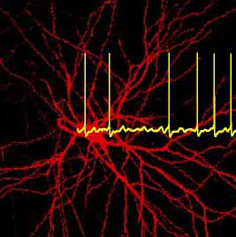
Neuron in the lateral nucleus of the
amygdala Source: Thomas Heinbockel, University of Arizona. |
|
These
observations suggest that the information that enters the
amygdala becomes represented at multiple locations within
it. Moreover, there seems to be a point-to-point correspondence
in the way this happens, so that the spatial organization
of the groups of neurons is preserved.
The integration of these various representations,
which originate in systems responsible for such widely differing
functions as long-term
memory, internal balance, and auditory perception, therefore
depends on the numerous internal connections among the various
nuclei of the amygdala. The evidence does seem to indicate
that these connections are sufficiently numerous, complex,
and diversified to play this role. |
Indeed, contrary to what was initially believed,
the information flow within the amygdala is highly reciprocal.
It does not simply travel in one direction, from the main entryway
at the lateral nucleus to the exit from the central nucleus. Indeed,
most of the main targets of the lateral nucleus send projections
back to it as well.
The representations thus encoded in the amygdala
and modulated by other brain structures ultimately converge at
the output areas–primarily the central nucleus and the amygdalohippocampal
area. This integration enables the brain to generate an activity
pattern that can trigger the appropriate changes in the various
structures responsible for the emotional reponse to the situation. |
|