Tool Module: Identifying Pathways in the Brain
To identify the connections between the various structures in the brain, researchers can work directly with the human brain, but often, they use animal models instead.
In animal studies, researchers can use two different types of experimental techniques.
(A) Degeneration techniques, based on the reaction of neurons to lesions
When an axon is disconnected from the body of its neuron, it degenerates and is eventually attacked by phagocytic cells. Researchers therefore produce highly localized lesions, then use dyes to trace certain pathways. This approach is limited, however, because not all neurons display marked degeneration after a lesion.
(B) Tracing techniques, based on the ability of neurons to transport molecules through their axons
To locate the origin of a neural pathway, researchers can inject a molecular marker such as the enzyme horseradish peroxidase into the region where the axons terminate. This marker is then transported toward the neuronal cell bodies by a phenomenon known as retrograde axonal transport. Once it reaches these cell bodies, the marker catalyzes a reaction whose product can be detected under an electron microscope. Thus the neurons where the pathway originates are revealed.
Conversely, researchers can also determine the point where axons terminate by injecting certain radioactive precursors into the region where the neuronal cell bodies are located. The cell bodies incorporate these radioactive precursors into macromolecules, which are then carried to the ends of the axons by anterograde axonal transport. Radiation-sensitive film is then used to capture images of the pathways thus marked.
Source: The Emotional Brain, by Joseph LeDoux
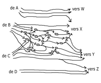
Even though the brain contains billions of neurons that form an incredibly complex network, such tracing techniques can reveal very specific connection patterns. In the diagram here, for example, the central neural network receives inputs from regions B and C, but not from regions A and D. This network also sends outputs to regions X and Y, but not W or Z. Also, region C communicates with region Y both directly and through the central network.
When studying human brains directly, researchers obviously cannot create lesions or inject markers, so they must use other approaches.
Instead, they take advantage of the fact that after a lesion occurs naturally in the brain, there are long distances over which the axons do not regenerate. The space created in the nervous system by the disappearance of these axons becomes filled with astrocytes that will leave scars where the pathways once were. By observing such tissue changes following very specific types of accidental lesions, researchers have accumulated reliable data on the connectivity of the human brain.
These data, combined with those from animal studies, have given scientists a good idea of the general connectivity of the brain. They also help us understand why we do not yet have a complete picture of all the pathways in the brain.