Experiment Module: How the Brain Keeps Information from the Left and Right Eyes Separate
In the early 1970s, neurophysiologists
David Hubel and Torsten Wiesel conducted a clever experiment to try to understand
how the separation of information from the right eye and the left eye was maintained
all the way to layer IV C of the striate cortex.
The researchers began
by injecting a radioactive amino acid into the eyeball of a monkey. This amino
acid was taken up by the ganglion cells of the retina, incorporated into proteins,
and then transported up the axons of these cells toward the lateral geniculate
nucleus (LGN). Some of these radioactive proteins then successfully crossed over
from the tips of the ganglion cell axons and were incorporated into the LGN cells
that processed visual information from the eye where the radioactive amino acid
had been injected. And from there, these proteins travelled up the axons of the
LGN cells and entered layer IV C of the primary visual cortex.
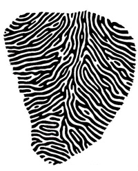
Hubel and Wiesel then took thin sections of the monkey's visual cortex and applied them to a radiation-sensitive film (a method called autoradiography). The resulting images showed areas that were coloured by clusters of silvery grains, indicating that these areas had received projections from the eye injected with the radioactive amino acid.
Source: Hubel and Wiesel,
1977.
In these tissue sections, Hubel and Wiesel observed
that the distribution of nerve endings from the injected eye was discontinuous
and took the form of regularly spaced bands which they termed ocular dominance
columns. In subsequent experiments, Hubel and Wiesel showed that the
spaces between the bands innervated by the left eye were indeed, as they had suspected,
innervated by the right eye. The researchers concluded that the nerve endings
of the left eye and the right eye in layer IV were arranged in alternating adjacent
bands, somewhat like the stripes of a zebra.
| |
![]()